A New Era in Breast Cancer Screening? AI-Powered Simulated Contrast-Enhanced Breast MRI Shows Promise
New technology could increase breast MRI accessibility, improve health outcomes


In concert with the RadioGraphics monograph, RSNA News shares the latest in breast cancer research, intervention and patient education.
Contrast-enhanced breast MRI, the most sensitive test for breast cancer detection, is recommended as annual supplemental breast cancer screening for high-risk patients, and some are using this technology in addition to yearly mammography to screen women with extremely dense breasts.
However, there are several potential barriers to expanding contrast-enhanced MRI screening to average-to-intermediate risk women, such as the time needed to obtain intravenous access, additional scanning time for post-contrast sequences, the cost of gadolinium contrast agent and the additional labor required—a physician must be present to monitor any adverse reactions to the contrast.
In addition, some patients are concerned about the unknown long-term effects of gadolinium deposition in the brain and bones. The use of gadolinium in pregnant patients or patients with end-stage renal disease is contraindicated, as gadolinium can cross the placenta, or trigger nephrogenic systemic fibrosis (NSF) in patients with impaired renal function.
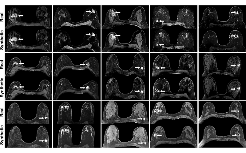
In a new Radiology proof-of-concept study, researchers evaluated the feasibility and accuracy of AI-simulated contrast-enhanced breast MRI, using precontrast and contrast-enhanced MRIs of patients with biopsy-proven invasive breast cancer. The simulated images demonstrated a high degree of similarity to clinical images, indicating that simulated contrast enhanced MRI shows promise as a potential tool to increase the accessibility of MRI screening.

Real versus simulated (ie, synthetic) contrast-enhanced T1-weighted axial breast MRI scans of patients with invasive breast cancer. Pairs of real and simulated contrast-enhanced breast MRI scans from 15 patients with invasive breast cancer (arrows). Intrathoracic and extramammary structures were masked in all images. https://doi.org/10.1148/radiol.213199 ©RSNA 2022
Proof-of-Concept Study Demonstrates Feasibility
The research study selected 96 patients with invasive breast cancer who underwent contrast-enhanced breast MRI exams at a single academic institution. The researchers utilized five precontast sequences to train a deep neural network to simulate contrast-enhanced MRI scans.
For qualitative assessment, four breast radiologists, blinded to whether the method of contrast was real or simulated, assessed image quality, presence of tumor enhancement, and maximum index mass size by using 22 pairs of real and simulated contrast-enhanced MRI scans.
The readers assessed all simulated scans as having the appearance of a real MRI scan with tumor enhancement, while index mass sizes on real and simulated scans demonstrated good to excellent agreement. Readers rated 95% of the simulated MRI scans to be of diagnostic quality.
The results indicate that it is feasible to generate simulated contrast-enhanced breast MRI scans with use of deep learning. Co-lead investigator Maggie Chung, MD, assistant professor of radiology, University of California, San Francisco, shared that the researchers were pleased with the results.
“We were pleasantly surprised that we were able to accomplish these results with a small pilot dataset of biopsy-proven invasive cancers,” she said. “It's very promising for our future work.”
Continued Study May Enable Technology to Detect Small Masses and Non-Mass Enhancement
In an accompanying commentary, Priscilla J. Slanetz, MD, MPH explained that the main limitations of this study are its small data set and focus on large masses.
“The goal of breast cancer screening is to find cancers under a centimeter in size, because those are going to have the best prognosis and are less likely to be axillary node positive,” said Dr. Slanetz, who is a professor of radiology at Boston University Chobanian & Avedisian School of Medicine, and a breast radiologist at Boston Medical Center. “It's a strong proof-of-concept study, but the technology must be able to work for small lesions.”
Dr. Slanetz added that simulated contrast-enhanced breast MRI must also be capable of detecting non-mass enhancements.
“Given that not all cancers present as a mass, such as ductal carcinoma in situ and some invasive cancers, it must be certain that this technique can detect non-mass enhancement as well,” she said.
Dr. Chung and her colleagues are optimistic that these limitations can be addressed with further study, using larger data sets and an improved deep learning model.
“In our pilot study, we were able to simulate enhancement in cancers as small as seven millimeters, so we are optimistic that we can improve upon this with a much larger data set and with an improved AI model,” Dr. Chung said. “Upon visual inspection, we did observe some simulation of non-mass enhancement and will further refine this in our follow-up study.”
“Simulated contrast opens up the possibility of offering MRI screening during pregnancy. Also, for pregnant women with breast cancer, simulated contrast MRI could be performed for evaluation of extent of disease to obtain important information for treatment planning sooner.”
MAGGIE CHUNG, MD
Increased Accessibility, Patient Compliance and Decrease In False Positives
If this technology were to one day have a high degree of reliability, it would present numerous patient benefits, including increased affordability. Dr. Slanetz hypothesized that lower-cost, simulated contrast-enhanced breast MRI exams could remove financial barriers for some patients.
“Breast MRI is a relatively high-cost test due to the administration of intravenous gadolinium, which is expensive,” Dr. Slanetz said. “For those patients who are not well-insured, there could be out-of-pocket costs that prohibit them from being able to access the technology.”
Dr. Chung explained that simulated contrast would also make this exam accessible to pregnant patients or patients with kidney disease.
“Simulated contrast opens up the possibility of offering MRI screening during pregnancy,” Dr. Chung said. “Also, for pregnant women with breast cancer, simulated contrast MRI could be performed for evaluation of extent of disease to obtain important information for treatment planning sooner.”
It is predicted that this technology would also improve health outcomes in a variety of ways, including patient compliance.
“Removing the need for IV placement and additional time in the MRI scanner could make the exam more comfortable and shorter, and help patients better tolerate the exam,” Dr. Chung said. “If patients are willing to have this exam every year, we improve our chances of finding cancers as early as possible and achieving better patient outcomes.”
It is also possible that this deep learning tool may help radiologists provide more accurate readings, thereby leading to better patient outcomes.
“It could help decrease some of the variability in radiologists’ interpretations, because the algorithm will help radiologists focus on what's important, and result in more consistency among readers,” Dr. Slanetz noted. “This technology, depending on how well it works, could help us reduce the number of false positives.”
Finally, simulated contrast-enhanced breast MRI would decrease radiologists’ workload—a very desirable goal in the midst of a breast imager shortage, according to Dr. Slanetz.
“One of the challenges we face right now is a relative shortage of breast imagers,” she said “If this was really a reliable tool, the image interpretation time for breast MRI could go down significantly. It could be that we won't necessarily need to look at images where the algorithm has not detected any areas of concern.”
For More Information
Access the Radiology study, “Deep Learning to Simulate Contrast-enhanced Breast MRI of Invasive Breast Cancer.”
Read previous RSNA News stories on breast imaging:
- Radiologists Play a Key Role in Assessing Radial Scars
- Understanding How Pregnancy and Breastfeeding Impact Breast Imaging and Diagnosis
- Enhancing the Evaluation of Breast Symptoms in the ED