Radiologists Play a Key Role in Assessing Radial Scars
New technology increases need to recommend management options

In concert with the RadioGraphics monograph, RSNA News shares the latest in breast cancer research, intervention and patient education.
Increased use of digital breast tomosynthesis (DBT) for mammographic screening has led to more frequent identification of architectural distortion (AD) and the potential for unnecessary surgery. Breast radiologists play an important role in management recommendations following screening and can provide guidance and imaging follow-up to help patients avoid overtreatment.
“An increase in discovery of architectural distortion leads to more biopsies, as one cannot assess whether the cause of the architectural distortion is due to a malignancy or a benign process solely based on imaging appearance,” said Ana P. Lourenco, MD, professor of diagnostic imaging in the Department of Diagnostic Imaging at the Warren Alpert Medical School of Brown University in Providence, RI.
According to Dr. Lourenco, radial scars and complex sclerosing lesions, often collectively referred to as radial sclerosing lesions (RSLs), are among the benign entities that are targeted for biopsy because of their suspicious imaging appearance. RSL imaging features can be detected on DBT and may overlap with breast malignancy. They can be found in isolation or associated with atypia or other high-risk lesions which have malignant potential.
“With the introduction of DBT into clinical practice, we are doing more biopsies of architectural distortions and therefore identifying more radial scars at biopsy,” Dr. Lourenco said. “The traditional management for radials scars has been surgical excision, but the likelihood of finding cancers in this scenario is extremely low in multiple newer studies.”
A RadioGraphics article co-authored by Dr. Lourenco and colleagues provides a review of the overall histopathologic, clinical and imaging features of RSLs and provides perspective on appropriate management based on currently available data regarding lesion upgrade rates.
Watch Dr. Lourenco discuss the role that radiologists' play in assessing radial scars:

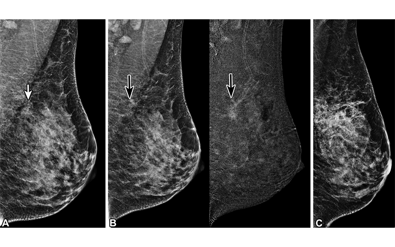
Complex sclerosing lesion (CSL) CSL in a 41-year-old woman. (A) Screening mediolateral oblique DBT mammogram of the left breast shows architectural distortion (arrow). The patient was recalled for contrast-enhanced mammography. (B) Mediolateral oblique contrast-enhanced mammogram (left) and recombined image (right) of the left breast shows nonmass enhancement (arrows, left) (C) Mediolateral digital mammogram shows the biopsy clip at the site of architectural distortion. DBT-guided core biopsy was performed, revealing CSL, which was confirmed at subsequent surgical excision. https://pubs.rsna.org/doi/10.1148/rg.230022 ©RSNA 2023
RSL Clinical Presentation and Imaging Manifestations
Dr. Lourenco and colleagues noted that RSLs are most commonly found in asymptomatic women between the ages of 30 and 60 years old, and are not usually palpable or associated with skin thickening or retraction. An RSL is a commonly occurring histopathological finding, an incidental microscopic finding on surgical excision specimens, or an incidental finding on mammography.
The authors evaluated and described imaging manifestations across a variety of modalities.
• Mammography
RSLs may be multiple and/or distributed bilaterally. Mammographic features are characterized by stellate or spiculated appearance with AD and central lucency. Less commonly, RSL may present as a mass or calcification. Because it is not possible to differentiate AD associated with cancers from AD associated with non-cancers on mammography, biopsy is often necessary and leads to increased diagnosis of radial scars. However, despite the increased detection of RSLs by DBT, one study referenced by the authors showed no difference in upgrade rates before and after the introduction of DBT.
• Ultrasonography
Frequently, RSLs are not visible on US. While there have been attempts to use techniques like elastography to differentiate RSLs and AD associated with breast cancer, RSLs may also exhibit inherent stiffness on elastography similar to that of breast cancer. The authors note that this suggests elastography cannot reliably differentiate RSL from malignancy.
• Contrast-Enhanced Magnetic Resonance Imaging
RSLs have a variable appearance on breast MRI, including as an irregular enhancing mass sometimes exhibiting AD, an enhancing focus, AD without associated mass, and non-mass enhancement. RSLs visible on MRI can demonstrate variable enhancement kinetics. The authors note that some centers use MRI to triage patients with biopsy-proven RSLs either to excision or surveillance.
• Contrast-Enhanced Mammography
Two studies referenced in the article looked at AD on contrast-enhanced mammography (CEM) and its association with RSLs. In one, there were no identifying features used to differentiate between benign and malignant enhancement and no data showing if CEM could be used to predict upgrade rates. Dr. Lourenco and colleagues note that more data are needed as the clinical use of CEM increases.
“Using techniques that decrease upgrade rates may allow patients to undergo imaging follow-up to monitor changes over time, rather than undergoing invasive surgical excision.”
ANA P. LOURENCO, MD
Management and Upgrade Considerations
RSL management practices are evolving and can incorporate a multidisciplinary approach that may include discussion of patient risk factors and allow for better patient-informed medical decision making as one decides whether to proceed to surgery or not.
“RSLs without atypia on biopsy of architectural distortion alone, without associated calcification or mass, may be appropriate for surveillance, particularly if the area of distortion is small,” Dr. Lourenco said. “However, there is no consensus regarding surveillance management protocols, and there is no evidence to suggest the most appropriate surveillance interval.”
According to the authors, prior research has shown that accurate image-guided biopsies with larger tissue samples yield lower upgrade rates. RSLs without associated atypia have very low upgrade rates at surgery.
“Using techniques that decrease upgrade rates may allow patients to undergo imaging follow-up to monitor changes over time, rather than undergoing invasive surgical excision,” Dr. Lourenco said.
For More Information
Access the RadioGraphics article, “Imaging and Management of Radial Scars and Complex Sclerosing Lesions."
Read previous RSNA News articles about breast cancer imaging: