3D Printed Models Move to the Head of the Class
Models provide invaluable education simulation that is transferable to patients
Editor's Note: This is one in a series of articles on current topics in 3D printing applications for radiology. Read the previous article in the series.
There has been a lot of news about the use of 3D printed models for surgical planning. Created using a patient’s imaging studies, these models allow surgeons to not only see, but also physically interact with anatomical representations of the areas they plan to operate on. But these imaging-based 3D models also play a major role in medical education.
“As the anatomical detail provided by imaging increases, it becomes significantly more difficult to build traditional training models,” said Adam Birosh, MD, a radiology resident at the University of Ottawa. “3D printing offers a solution to this by allowing very complex models to be produced quickly and at low cost.”
“3D models are playing an increasingly important role in teaching anatomy to pre-med and medical students, and to provide more specialized training and skills to residents,” said Gerald Jit Shen Tan, MD, a consultant in diagnostic radiology at Tan Tock Seng Hospital in Singapore.
“In medical school, this technology has opened the door to a new generation of simulators, allowing students to practice procedures and techniques that had traditionally been taught using low fidelity models or learned ‘on the job,’” added Michael O’Reilly, MD, a consultant radiologist at the University of Limerick Hospital group in Ireland.
One of the key benefits of using these models is the provision of patient-specific cases, such as anatomical variations that could make otherwise routine procedures more challenging. As many of these procedures are done with imaging guidance, it is essential that the finished models look like what imaging revealed — which is where radiologists come in.
The Modern Anatomists
According to Dr. O’Reilly, radiologists are the modern anatomists.
“Many procedures that had traditionally been performed without imaging guidance or via anatomical landmarks — such as vessel access, chest drain placement, lumbar punctures and biopsies — are now performed using imaging guidance to help,” he explained. “If the models we create are not realistic when using ultrasound or fluroscopy, then they are not going to be of much help to the trainee.”
“Our key role is to select suitable scans for the use case and provide the raw digital data for printing,” Dr. Tan added. “In some institutions, such as mine, radiologists are also engaged in the actual training, particularly at the undergraduate level.”
Here, Drs. O’Reilly, Tan and Birosh, among others, have developed 3D printed models specifically for teaching purposes. For example, while teaching anatomy at University College Dublin, Dr. O’Reilly used a printed model of the bones and muscles of the lower limb that could be assembled like a 3D puzzle.
“Much anatomy is taught by taking things apart, but a lot of the learning occurs when we force people to try to put it back together,” Dr. O’Reilly said.
He also helped create a model for teaching ultrasound and fluoroscopically guided femoral artery access to trainees who perform these procedures (e.g., interventional radiology, cardiology and vascular surgery residents).
“We developed this model with experienced interventional radiologists, who helped us realize that, to make the model realistic and challenging enough for trainees to want to use it, we needed to include the femoral head and the femoral vein, and to have the vessels pressurized,” Dr. O’Reilly added.
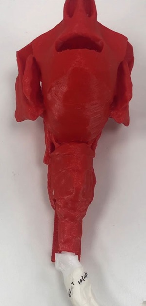
Dr. Tan, on the other hand, was involved in printing 3D models of the bronchi. The life-sized models are based on CT scans showing pathologies such as a bronchial tumor or external compression by an enlarged thyroid. To ensure the models mimicked the texture and feel of the human airway with fidelity, they were printed using a unique blend of hard and soft materials.
“These models allow bronchoscopists to train on more complex pathologies that are not available with standard simulators, at a reasonable price and with the potential to customize the model to individual patients,” Dr. Tan explained.
Dr. Tan noted that, with the bronchial model working well as a proof-of-concept, it can now be expanded to other anatomical regions and specialties, such as for nasoendoscopy in head and neck surgery.
Meanwhile, Dr. Birosh helped develop a 3D model of the human arterial system that ranges from the base of the aortic arch up beyond the circle of Willis. The model, which is used in training for neurovascular access procedures, allows trainees to insert a catheter into the base of the model and navigate it into the cerebral arteries, just as they would with a real patient.
According to Dr. Birosh, one big advantage of this model is it can be used with or without fluoroscopy.
“Typically, the only way to see the catheter and wire in the patient is by using live fluoroscopic imaging to track the movement through the vessels, which can be difficult since conventional fluoroscopy only provides a two-dimensional image,” Dr. Birosh explained. “This model allows you to practice navigating the catheter in a fluoroscopy lab, just by looking at the catheter through the clear walls of the vessels.”
An example of a specific procedure that requires cerebral vascular access is endovascular thrombectomy (EVT), in which clots are retrieved from cerebral arteries to treat strokes. The speed at which a clot is removed from the artery will affect how much brain tissue is affected by the lack of blood flow, and how severely.
“Being able to practice this procedure prior to performing it on real patients could greatly improve the confidence of the radiologist going into the procedure,” Dr. Birosh added.
Improving Skill Levels
At the Hospital for Sick Children, which is affiliated with the University of Toronto, 3D printed heart models are being used to provide training in congenital heart surgery. For example, the Hands-On Surgical Training (HOST) platform is designed to provide surgeons the opportunity to practice procedures on various congenital heart defects using simulation with 3D printed models.
The hospital offers a 2 1/2 day HOST course, where surgeons are coached by experienced congenital cardiac surgeons using the 3D printed heart model. They’ve also incorporated a monthly HOST program as part of its congenital heart surgery curriculum for surgical fellows.
According to Shi-Joon Yoo, MD, professor of medical imaging at University of Toronto, and head of cardiac imaging at the Hospital for Sick Children, where he is also clinical director of the 3D printing program, these courses aim to provide the surgeons with ample opportunity to practice various surgical procedures without risking patient safety.
“We also provide our physicians with additional opportunities to learn congenital heart morphology by directly observing pathologic specimens and 3D printed models,” Dr. Yoo added. “There is no doubt that anatomically correct 3D printed models are great resources for improving surgical skills.”
"In medical school, this technology has opened the door to a new generation of simulators, allowing students to practice procedures and techniques that had traditionally been taught using low fidelity models or learned ‘on the job.’"
Michael O'Reilly, MD,
More Opportunity Ahead
While these are all excellent examples of 3D printing being used as a teaching tool, according to Dr. Birosh, they only scratch the surface of what’s possible.
“In general, 3D printing has been greatly underutilized for education simulation,” he said. “While some research articles have demonstrated potential applicability, there are very few examples of training programs actually implementing these tools for regular resident, student or fellow procedural training.”
But with the complexities of 3D printing technology increasing — and the costs of 3D printing decreasing — the pendulum could soon start to swing the other way.
“As the technology improves, it becomes easier to develop accurate 3D replicates of more areas of the human body, making these models even more useful as teaching tools,” Dr. Birosh said. “Furthermore, any decrease in production costs will improve program uptake and accessibility of the training tool.”
As more 3D printed models find their way into training programs, the net result will be better patient care.
“Patients with atypical and challenging anatomy that is associated with higher complication rates often don’t present commonly enough for adequate teaching,” Dr. O’Reilly concluded. “3D printing allows us to ‘print’ the difficult cases so trainees can get near hands-on experience and then take this knowledge — and confidence — into their practice.”
For More Information
Learn about the RSNA 3D Printing SIG.
Read previous RSNA News stories on 3D printing:
- 3D Printed Models Move Complex Surgery in Exciting New Directions
- Defining Radiology’s Role in the 3D Printing Explosion
Watch RSNA videos with information on 3D printing: