Imaging at the Heart of Sports Cardiology
Radiologists play key role in risk assessment and return-to-play decisions

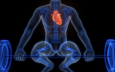
As participation in competitive and recreational sports continues to grow, so does recognition of sports cardiology as a critical, rapidly evolving subspecialty. At the intersection of cardiovascular health and athletic performance, this field demands precise, nuanced assessment—particularly when lives and careers may hinge on the difference between imaging features related to benign adaptation and other pathological conditions.
A RadioGraphics article by Prabhakar Shantha Rajiah, MBBS, MD, a radiologist at Mayo Clinic in Rochester, MN, and colleagues outlines how advanced imaging techniques—particularly cardiac MRI (CMR) and coronary CT angiography (CCTA)—support accurate diagnosis and risk assessment in sports cardiology.
“Multimodality imaging is an essential tool in the sports cardiologist’s arsenal,” Dr. Rajiah said. “It provides critical support for risk assessment and differential diagnosis within a multidisciplinary framework.”
Distinguishing Disease from Adaptation
A major challenge in sports cardiology is distinguishing between physiologic adaptations to intense training—commonly known as athlete’s heart—and underlying cardiovascular disease. Misinterpretation in either direction can have significant consequences.
“A primary goal during pre-participation screening and evaluation of athletes is to differentiate between physiological adaptations and true pathology,” Dr. Rajiah said. “Mislabeling normal adaptation can unnecessarily sideline an athlete, while missing a pathological finding can have serious implications.”
Pre-participation screening typically includes clinical history, physical examination and electrocardiography (ECG), with exercise ECG added for older athletes. While echocardiography is often the first imaging step with abnormal initial assessment, advanced imaging becomes essential when echocardiographic findings are equivocal or raise concerns for more serious underlying pathology. In such cases, CMR and CCTA are key next steps.
“CMR is particularly useful when echocardiographic findings are inconclusive, helping to differentiate athlete’s heart from mimicking pathologies,” Dr. Rajiah said. “Additionally, CMR supports diagnosis and risk stratification in symptomatic athletes or those recovering from significant cardiac events.”
CMR is particularly valuable in borderline cases, as the tissue characterization capabilities of MRI can provide essential information that either complement or extend beyond echocardiographic findings. “Diagnosis takes into account clinical pretest probability, sport type, competition level, age, sex, body size, ECG and exercise test findings, and imaging results,” Dr. Rajiah explained.
These imaging modalities can also guide decisions related to sports eligibility and long-term surveillance. “CCTA offers high-resolution evaluation of coronary artery anomalies and coronary artery disease (CAD), making it an invaluable component in certain clinical scenarios,” Dr. Rajiah added.
In trained athletes, findings such as symmetric left ventricular hypertrophy, mild chamber dilation and preserved or slightly reduced ejection fraction often reflect benign adaptation. However, similar findings in different clinical contexts may suggest cardiomyopathies, key differential diagnoses. CMR helps mitigate the ambiguity by precisely assessing these features alongside myocardial tissue characteristics, thereby supporting the exclusion of cardiovascular disease in the appropriate clinical context.
“CMR is also valuable in evaluating athletes with symptoms such as palpitations or chest pain, or in those recovering from events like arrhythmia, myocardial infarction or resuscitated cardiac arrest,” Dr. Rajiah added.
Balancing Risk, Access and Imaging Use
CAD is a rare but important consideration in athletes presenting with chest pain, especially master athletes. CCTA can be a strong first-line test in those cases, Dr. Rajiah said. By providing high-resolution visualization of coronary anatomy, CCTA is the preferred non-invasive method for the evaluation of coronary anomalies.
Karen Ordovas, MD, MS, professor of radiology and chief of cardiothoracic imaging at the University of Washington in Seattle, emphasized the importance of interpreting imaging findings within the context of exercise-induced remodeling.
“The key for radiologists is to understand how different kinds of strenuous exercise can lead to changes in the heart that could be confused with diseases,” she said. “Understanding the expected changes that can happen due to endurance versus resistance training can be key when interpreting CMR in athletes.”
Dr. Ordovas noted that while CMR is a powerful tool, it is not universally accessible. “There are still rural and some suburban areas in the U.S. where access to CMR remains limited,” she said.
During the COVID-19 pandemic, Dr. Ordovas co-authored a consensus statement on CMR use in athletes recovering from infection. “There was a surge in requests for CMR to ‘clear’ athletes to return to play, but imaging should not be used as a blanket screening tool,” she said. “CCTA and CMR are best reserved for cases with elevated risk or persistent cardiac symptoms.”
“The key for radiologists is to understand how different kinds of strenuous exercise can lead to changes in the heart that could be confused with diseases. Understanding the expected changes that can happen due to endurance versus resistance training can be key when interpreting CMR in athletes.”
Karen Ordovas, MD, MS
Evolving Field, Expanding Role for Imaging
As sports cardiology matures into a recognized subspecialty, imaging is increasingly central to its evolution. “Sports cardiology is a dynamic and rapidly evolving specialty,” Dr. Rajiah said. “As our understanding deepens, recommendations for sports participation and return-to-play after cardiac events are continually refined.”
Both Dr. Rajiah and Dr. Ordovas emphasized the importance of radiologists participating directly in clinical discussions.
“Sports cardiology requires a specialized understanding of how exercise-induced cardiac remodeling presents on imaging,” Dr. Rajiah said. “It also requires familiarity with the physiologic demands of various sports and how underlying pathologies may interact with those demands.”
To make accurate and meaningful contributions, radiologists must embed themselves in the clinical process, working closely with cardiologists, team physicians and athletic trainers.
“It’s not just about what the image shows,” Dr. Rajiah added. “It’s about how that finding fits into the bigger picture and understand the overall process of evaluation and recommendations to help put imaging findings in the appropriate context.”
For More Information
Access the RadioGraphics article, "Utility of MRI and CT in Sports Cardiology."
Access the Journal of Cardiovascular Magnetic Resonance consensus paper.
Read previous RSNA News stories on cardiac imaging: