Open-Source AI Model for MRI Segmentation Receives Margulis Award
New tool extends TotalSegmentator’s capabilities to MRI, offering faster, more consistent anatomic analysis


Research developing an open-source, easy-to-use automatic segmentation model for MRI images has earned the 2025 Alexander R. Margulis Award for the best original scientific article published in Radiology.
“This study represents a major advance in medical image analysis,” said Jacob Sosna, MD, chair of the Margulis Award for Scientific Excellence Selection Committee and the RSNA Publications Council. “By developing a segmentation algorithm that operates independently of specific MRI sequences, the researchers overcame a key limitation of traditional methods that require sequence-specific training. This sequence-agnostic design enhances the robustness and clinical versatility of automated segmentation across diverse MRI protocols—bringing the field closer to reliable, scalable AI tools that can be applied universally in radiology.”
MRI plays an essential role in diagnosing a wide range of medical conditions throughout the body. However, manual segmentation of MRI scans remains one of the most time-consuming and tedious tasks, often requiring hours of expert work.
The self-configuring framework nnU-Net, a type of AI model designed to automatically recognize and outline structures in images, has set new standards in medical image segmentation by adapting to any new dataset with minimal user interventions. Tools such as TotalSegmentator have already demonstrated success in enhancing the capabilities of nnU-Net on CT images.
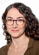
“While TotalSegmentator CT had become a widely adopted tool for robust automated segmentation of CT scans, there was no equivalent solution for MRI,” said lead author Tugba Akinci D'Antonoli, MD, a neuroradiology fellow from the Department of Diagnostic and Interventional Neuroradiology, University Hospital Basel, Switzerland. “We set out to create a tool that could bring the same reliability, simplicity and open-source availability to MRI.”
“Training the model with both CT and MRI data actually improved MRI segmentation performance. This suggests that CT data can serve as a form of data augmentation, helping the model generalize better to MRI.”
— TUGBA AKINCI D’ANTONOLI, MD
Building a Universal Model
To develop the tool, the researchers trained the nnU-Net-based model on a diverse set of major anatomic structures using 616 MRI and 527 CT images. The MRI datasets varied in contrast, section thickness, field strength and pulse sequence.
Eighty anatomic structures were identified for segmentation. The model was trained on MRI and CT images with 1.5-mm isotropic resolution, while a second model—TotalSegmentator MRI-3—was trained on images with 3-mm isotropic resolution. To manage the computer memory requirements needed to efficiently handle this training, the 80 anatomic structures were divided into six models.
TotalSegmentator MRI matched expert-drawn segmentations closely, showing strong accuracy across all 80 structures in the internal test set. For the 50 most clinically important structures, TotalSegmentator MRI was even more accurate. The model outperformed the publicly available MRSegmentator and AMOS on all comparable benchmarks.
Thanks to a diverse MRI training dataset, the model achieved consistent performance across a wide range of scan types, despite the inherent variability of MRI data. “Small vessels and low-contrast organs remain challenging, even for human experts,” Dr. D’Antonoli noted.
An unexpected finding emerged when combining CT and MRI data.
“Training the model with both CT and MRI data actually improved MRI segmentation performance,” Dr. D’Antonoli said. “This suggests that CT data can serve as a form of data augmentation, helping the model generalize better to MRI.”

Axial MRI images from the cases with the lowest (top) and highest (bottom) Dice score in the CHAOS external test set for our proposed model, TotalSegmentator MRI, as well as for two publicly available baseline models, MRSegmentator and AMOS. The reference segmentation for liver and spleen is shown in green, and the segmentation of the model is shown in red. The CHAOS dataset was used to show the best and the worst results because this dataset is the most independent from the training data of the three models. The CHAOS Challenge dataset is available at http://zenodo.org/records/3431873.
https://doi.org/10.1148/radiol.241613 © RSNA 2025
From Research to Real-World Application
To demonstrate clinical potential, the researchers applied TotalSegmentator MRI to a large internal dataset of 8,672 abdominal MRI scans to analyze age-related changes in organ volumes. They observed expected patterns, such as declining kidney, liver and spleen volumes with age, and increasing adrenal gland volumes.
“These studies can uncover clinically relevant insights, such as age-related changes in organ volumes that would otherwise be impractical to assess manually,” Dr. D’Antonoli said.
According to Dr. D’Antonoli, the model directly impacts clinical workflow.
“It reduces the manual workload for radiologists, minimizes inter-reader variability and accelerates processes like organ volumetry, treatment planning and opportunistic screening,” Dr. D’Antonoli said. “For patients, this translates into more accurate diagnoses, faster turnaround times and the possibility of large-scale population studies.”
Open Science Drives Innovation
By releasing the full model, training data and annotations, the researchers hope to mark an important milestone in the evolution of AI-driven image analysis.
“Open science is the cornerstone of scientific progress,” Dr. D’Antonoli emphasized.
The team is already seeing widespread community engagement, with new MRI segmentation tools emerging almost weekly, many of which are benchmarking against TotalSegmentator MRI.
Next steps include expanding anatomical coverage to include finer structures, such as peripheral vessels and small muscle groups, to support broader adoption in clinical practice.
The Margulis Award is given each year in honor of the late Alexander R. Margulis, MD, a distinguished investigator and inspiring visionary in the science of radiology and longtime chair of the Department of Radiology at the University of California, San Francisco. The Margulis Award will be presented to Dr. D’Antonoli during RSNA 2025 in Chicago, Nov. 30-Dec. 4.
“In the long term, we envision automated segmentation becoming as standard and invisible as spellcheck in word processors—something always running in the background, quietly improving precision medicine and patient care,” Dr. D’Antonoli concluded.
For More Information
Access the Radiology study, “TotalSegmentator MRI: Robust Sequence-independent Segmentation of Multiple Anatomic Structures in MRI.”
Read the RSNA News story about the 2024 Margulis Award recipient.