Top Images in 'Radiology' Showcase Dramatic Views of Cutting-Edge Imaging Techniques
Unexpected presentations, striking appearances and unique views offer educational opportunities for radiologists
Selections have been made for the 2023 Images in Radiology, an online medical imaging collection that is part of the journal Radiology. Each year, Images in Radiology publishes captivating images demonstrating important medical diagnoses and state-of-the-art medical imaging technology. These images exemplify the significant contributions made by radiology to the field of medicine.
Images in Radiology is the second most popular manuscript submission type, after original research submissions, with more than 500 cases submitted between Jan. 1, 2023, and Oct. 31, 2023. With the conclusion of 2022–2023 academic year, the Radiology In Training editorial board has reviewed the Images in Radiology collection published from July 2022 through June 2023 for the 2023 Top Images designation. Thirty eligible publications have been ranked on the basis of visual appeal, novel application or development of an imaging technique, and educational value.
The top three images, second runner-up, first-runner-up and winner, were selected by the Radiology in Training editorial board based on aggregate scores from individual editorial board members’ rankings. The images were selected based on three criteria: novel technology or unusual pathology, educational or thought-provoking and visually compelling.
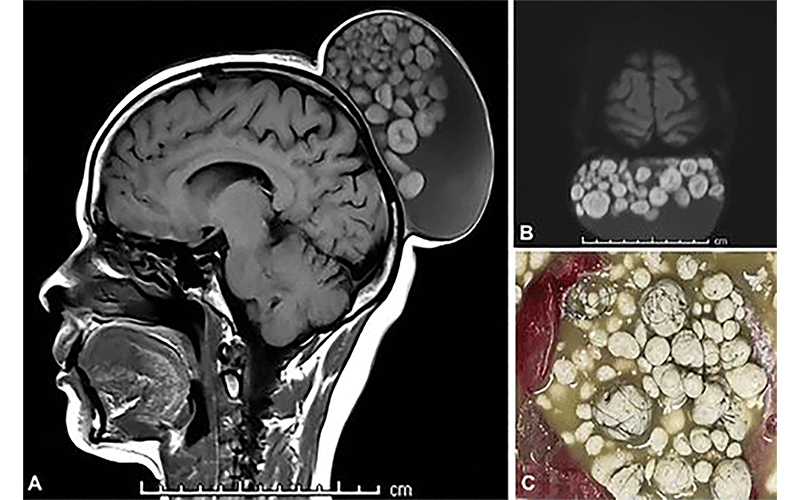
The 2023 Top Images in Radiology winner is entitled “Sack of Marbles Appearance of a Scalp Teratoma” by Sumit Thakar and Pavan Vasoya (1). The radiologicpathologic correlation of sebum-like material found in mature cystic teratomas, depicted on the MR and gross specimen images (Fig 1), is visually impressive, eye-opening, and educational. The illustration of this somewhat common entity in a less-common location embodies the core principles of radiology as a macropathology diagnostic specialty.

Images in a 52-year-old woman presenting with a scalp mass show a large cystic lesion in the subgaleal plane of the scalp with a “sack of marbles” appearance. (A) Sagittal unenhanced T1-weighted MRI scan shows a smooth-walled hypointense cyst with multiple spherical nodules that had isointense to hyperintense rims and thin hypointense cores. (B) Diffusion MRI scan shows the nodules demonstrating restricted diffusion. (C) Photograph of the gross specimen shows a sebumlike material within the cyst, hard spherical nodules, and strands of hair. https://doi.org/10.1148/radiol.230033 ©RSNA 2023
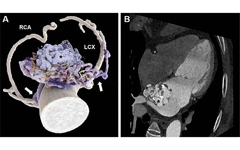
The first runner-up for this year’s Top Images in Radiology is entitled “Cinematic Rendering of Complex Coronary Artery Left Atrial Fistula” by Mingxi Liu and Xiaojuan Guo (2). The axial images showing multiple sinuses draining into the left atrium from the coronary arteries, although striking, may not allow for easy tracing of the vasculature. The crisp images postprocessed with cinematic rendering (Fig 2), however, carefully depict this unusual entity of complex coronary artery left atrial fistula and help the interpreting radiologists reach the final diagnosis.

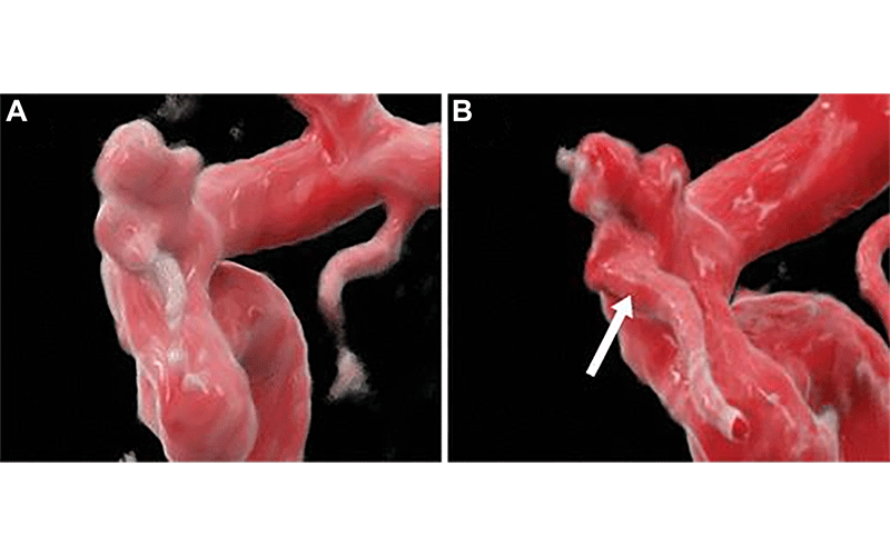
The second runner-up for the 2023 Top Images in Radiology is the publication entitled “Cerebral Aneurysm Imaging at 7T with Use of Compressed Sensing” by Amit Desai and Erik Middlebrooks (3). The authors’ direct comparison of the MR angiogram time-of-flight assessments at 7T versus 1.5T of an intracranial aneurysm (Fig 3) is an illustrative example of the incremental benefit that higher-field-strength MRI can offer, when available.

The other articles ranked among our top 10 images for the academic year, in alphabetical order, included:
• “Anomalous Origin of the Right Pulmonary Artery from the Aorta” (4)
• “CT-based Age Estimation of a Mammoth Tusk” (5)
• “High-resolution (7-T) Liver MRI for Pathologic Examination” (6)
• “Idiopathic Mesenteric Phlebosclerosis” (7)
• “Kawasaki Disease with Unusual Systemic Arterial Aneurysms” (8)
• “Thoracic Outlet Syndrome Associated with a Rib Synostosis” (9)
• “Total Anomalous Pulmonary Venous Return” (10)
For More Information
Access the Radiology article, “2023 Top Images in Radiology: Radiology in Training Editors’ Choices.”
Review the 2022 Images in Radiology winners.
Review the 2021 Images in Radiology winners.