RSNA Radiology Journal Recognizes Its Top Images of 2021
Radiology In Training editors’ select the top three images of the year
The top images of 2021 featured in Images of Radiology, an online medical imaging collection that is part of the journal Radiology, have been selected. Each year, Images in Radiology publishes compelling images that demonstrate important medical diagnoses and state-of-the-art technology in radiology. These images exhibit the unique contributions of radiology to the field of medicine at large.
This year, twenty-eight research images, published between July 1, 2020, and June 30, 2021, were eligible. The top three images were selected by the Radiology In Training editorial board members, a community of trainees who engage with the Radiology editorial board as editors, reviewers, authors and readers.
The images were selected based on three criteria: novel technology or unusual pathology, educational or thought-provoking and visually compelling. The board selected one winner and two runners-up.
The winner selected as the top 2021 Image in Radiology is from the article entitled “Micro-CT of Early Human Development” by Yousif Dawood and Bernadette S. de Bakker. This article received the highest ranking for showcasing cutting-edge imaging technology that could capture the fascinating beauty of early human development and emphasized the potential role of imaging in many areas of clinical and bench research.
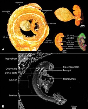
(A) Contrast-enhanced micro-CT at 3-µm isotropic resolution shows an intact ectopic pregnancy in the fallopian tube with a 3-mm-long human embryo of 6-weeks gestation. Left: volume rendering of the micro-CT images shows the embryo and its yolk sac completely surrounded by trophoblast and fallopian tube. Right: the embryo (with developing organs) and yolk sac can be clearly seen (top) and it shows full resemblance with the stage 12 specimen presented in an atlas (bottom). (B) Sagittal 3-µm micro-CT image of a human embryo at 6-weeks gestation with isotropic resolution. The embryo is surrounded by trophoblast and covered by amnion. On the dorsal side, the somites and otic vessel are easily distinguished, whereas ventrally, the (blood-filled) heart tube is prominent. Radiology 2020; 297:32 ©RSNA, 2020
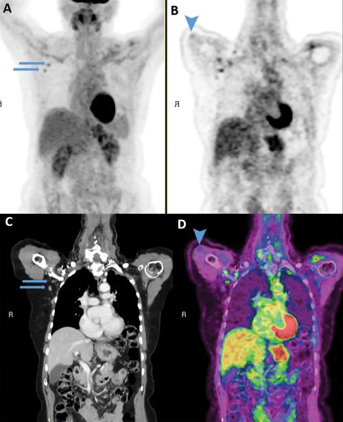
The first runner-up is an article entitled “Imaging of COVID-19 Vaccination at FDG PET/CT” by Michal Eifer and Yael Eshet. This article has been downloaded more than 24,000 times and, having been noticed and disseminated by the lay press, received more attention in online news and social media than 95% of all research publications scored by Altmetric.
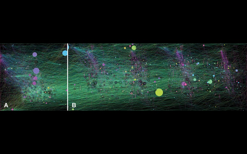
The second runner-up is “Radiologist Mouse Movements at a PACS Workstation” by Jan Vosshenrich and Hanns-Christian Breit. Although not based on a patient, these images speak to the human-computer interaction that is now core to the practice of radiology. The extensive and sometimes repetitive patterns, the authors suggested, point to a need for technologic solutions and attention to ergonomic design to optimize a radiologist’s workflow.
For More Information
Access the Radiology article, “2021 Top Images in Radiology: Radiology In Training Editors’ Choices.”
Review the 2020 Images in Radiology winners.