Low-Dose CT as Effective as Full-Dose CT Scans in Screening for COPD
Radiology: Cardiothoracic Imaging study shows promise for catching severe, chronic conditions earlier

The third leading cause of death in the U.S., chronic obstructive pulmonary disease (COPD) affects more than 15 million Americans and is often tied to emphysema and several genetic risk factors. While CT lung densitometry (CTD) is increasingly used to monitor progression and evaluate the efficacy of COPD treatment, variability of radiation dose levels and breath-hold volume between scans can reduce CTD reproducibility.
A promising new Radiology: Cardiothoracic Imaging study shows the effectiveness of low-dose CT imaging, used to screen for lung cancer, in screening for COPD and emphysema.
The study, led by Charles R. Hatt, PhD, an adjunct research professor of radiology at the University of Michigan, demonstrated that lung density measurements from low-dose CT scans can be used to identify clinical outcomes in patients with COPD just as effectively as from standard full-dose CT scans. And when some filtering is applied, low-dose CT scans can be just as useful at detecting real changes in disease progression as full-dose CT scans, results show.
“These findings should give physicians the confidence that lung density measurements from low-dose CT scans are just as effective in terms of prediction of outcomes as full-dose scans,” Dr. Hatt said. “Hopefully, that means we can do a better job of catching these severe, chronic diseases a little bit earlier.”
Results are especially critical considering that low-dose CT lung cancer screening is covered by the Centers for Medicare & Medicaid Services. And in 2021, the U.S. Preventive Services Task Force (USPSTF) updated its recommendation for annual screening for lung cancer with low-dose CT to include a broader patient base, expanding the recommended age range to 50 to 80 years (previously 55 to 80 years) of current and some former smokers, and reducing the pack-year history to 20 pack-years of smoking (previously 30 pack-years).
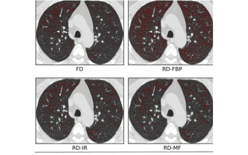
Effects of varying dose and noise reduction filtering. In this example, the difference in lung volumes between reduced-dose (RD) and standard fixed-dose (FD) scans were negligible and volume adjustment was not applied. Low-attenuating area less than −950 (LAA−950) differences between the FD, RD, RD-iterative reconstruction (RD-IR), and RD-median filter (RD-MF) scans presented in this study.
Hatt et al, Radiology: Cardiothoracic Imaging 2021 © RSNA 2021
COPDGene® Data Aids Understanding of Genetic Factors In this retrospective analysis of prospectively acquired data, Dr. Hatt and colleagues relied on the multicenter observational COPDGene® study designed to identify genetic factors associated with COPD.
Introduced in 2010, the COPDGene study comprises more than 10,000 subjects at 20 clinical sites throughout the country who undergo regular chest CT scans and provide blood samples and other clinical information.
Dr. Hatt, who has worked with the COPDGene study for the past six years, said the goal of his research is to better understand the genetic factors around the disease with the aim of improving quality of life for patients.
In the Radiology: Cardiothoracic Imaging study, Dr. Hatt and colleagues analyzed 1,205 patients enrolled in COPDGene who underwent full- and low-dose CT image acquisition protocols between November 2014 and July 2017.
The low-dose CT scan used about a quarter of the amount of radiation as a standard full-dose CT scan — the same low-dose scan used in the National Lung Cancer Screening Trial that enrolled more than 50,000 current or former heavy smokers. Dr. Hatt and colleagues also sought to determine if radiologists could detect emphysema and measure change in disease progression from the low-dose CT scans. Researchers wanted to determine the overestimation level and whether anything can be done to correct for that difference.
“These findings should give physicians the confidence that lung density measurements from low-dose CT scans are just as effective in terms of prediction of outcomes as the full-dose scans.”
Charles R. Hatt, PhD
Reducing “Noise” on Images
Dr. Hatt and colleagues analyzed the low-dose CT scans of 640 participants using iterative reconstruction such as applying median filtering to reduce noise on the images.
“Median filtering did an excellent job in terms of bringing us to what we would expect from a full-dose scan,” Dr. Hatt said. “And because median filtering is not proprietary, it can be widely implemented using post-processing software. So, anyone who has the right software can take a low-dose scan, run it through the median filter, and get similar results.”
And those results were promising — and often surprising, Dr. Hatt said.
“I was surprised that such a simple method could be used to harmonize the scan so well,” Dr. Hatt said.
Based on those findings and the predictive capacity of the low-dose scans, Dr. Hatt is confident that low-dose CT imaging can be used to provide a wealth of information beyond information obtained in lung cancer screening.
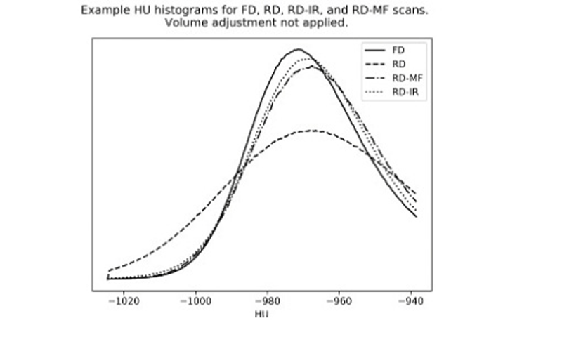
Effects of varying dose and noise reduction filtering. In this example, the difference in lung volumes between reduced-dose (RD) and standard fixed-dose (FD) scans were negligible and volume adjustment was not applied. Lung attenuation histograms for FD, RD, RD-MF, and RD-IR. FBP = filtered back projection.
Hatt et al, Radiology: Cardiothoracic Imaging 2021 © RSNA 2021
“While patients are getting scanned for lung cancer, we should be using that information to look for many other conditions, including COPD, emphysema, heart disease and bone disease,” Dr. Hatt said.
Dr. Hatt cited a study that demonstrated the effectiveness of low-dose CT in identifying emphysema in patients with and without a prior COPD diagnosis.
In the 2021 Clinical Imaging study, David Steiger, MD, and colleagues reviewed a prospective cohort of 52,726 patients who underwent baseline low-dose CT screening for lung cancer from 2003 to 2016 in the International Early Lung Cancer Action Program.
Emphysema was identified in 23.8% of patients undergoing low-dose CT and was unsuspected in 76.5% of patients, according to results.
Over time, Dr. Hatt said artificial intelligence (AI) software could also help automate processes to provide as much information as possible from one type of scan. “That is going to improve patient care and allow us to catch these diseases earlier,” Dr. Hatt said.
For More Information
Access the Radiology: Cardiothoracic Imaging study, “Comparison of CT Lung Density Measurements between Standard Full-Dose and Reduced-Dose Protocols.”
Access, “USPSTF Issues New Lung Cancer Screening Recommendations.”
Read previous RSNA News articles on low dose CT: