Low-dose Chest CT Leaves DNA Intact
Results help allay fears that screenings will lead to an increase in radiation-induced cancer

The low-dose chest CT scans used in lung cancer screening do not appear to damage human DNA, according to a study appearing in Radiology.
Low-dose CT screening for lung cancer in high-risk patients such as longtime smokers gained favor after the National Lung Screening Trial. The trial reported that use of low-dose chest CT scans could significantly reduce deaths from lung cancer compared to screening with chest X-rays. CT was able to identify cancers at an earlier, more treatable stage.
Along with the promise of CT screening came worries over the effects of radiation exposure on patients, as even the low-dose exam delivers more radiation than an X-ray—radiation that could affect DNA and potentially lead to cancer. Studies of these potential effects that rely on epidemiology have limitations, according to study senior author Satoshi Tashiro, MD, PhD, director of the Research Institute for Radiation Biology and Medicine at Hiroshima University in Hiroshima, Japan. A biological approach that looks at the effects of exposure on DNA has more power, he said.
“The National Lung Screening Trial suggested the value of low-dose CT screening in high-risk population for developing lung cancer,” he said. “There were, however, no studies investigating the biological effect of low-dose CT scans on large numbers of patients. These findings led us to investigate these effects.”
Results Show DNA Differences Between Standard- and Low-Dose CT
Dr. Tashiro and colleagues developed a system to look for damage and abnormalities in chromosomes. In a previous study, the technology showed increases in chromosomal aberrations after standard CT scans.
For the new study, the researchers compared the DNA in 107 patients who underwent low-dose chest CT with that of 102 who had standard-dose chest CT. They obtained blood samples before and 15 minutes after CT. The median effective radiation dose of low-dose CT was 1.5 millisieverts (mSv). The standard CT dose was 5.0 mSv.
Analysis of the DNA found significant differences between the group that had a standard-dose chest CT scan and those who had a low-dose chest CT.
“We could clearly detect the increase of DNA damage and chromosome aberrations after standard chest CT,” Dr. Tashiro said. “In contrast, no significant differences were observed in these biological effects before and after low-dose CT.”
Although low-dose CT is now commonly used for screening exams, standard CT is an effective diagnostic tool that is appropriate when the benefits outweigh any potential risk.
While the study did not endorse lung cancer screening with low-dose CT, its results appear to ease concerns over a potential increase in radiation-related cancer risk related to screening programs.
“Even using these sensitive analyses, we could not detect the biological effects of low-dose CT scans,” Dr. Tashiro said. “This suggests that application of low-dose CT for lung cancer screening is justified from a biological point of view.”
Beyond lung cancer screening, Dr. Tashiro said the DNA analysis could be used to study the biological effects of other types of imaging.
“We are interested in the biological effects of various types of radiological diagnosis, including PET/CT, to establish a better system for the management of medical radiation exposure,” he said.
For More Information
Access the Radiology study, “Biological Effects of Low-Dose Chest CT on Chromosomal DNA."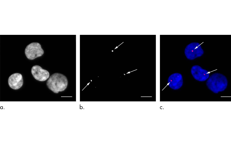
Immunofluorescent staining performed to depict γ-H2AX foci. Representative images of γ-H2AX foci in peripheral blood lymphocytes in an 82-year-old woman who underwent standard-dose CT. (a) Nuclear DNA of four lymphocytes. (b) γ-H2AX foci (arrows). (c) Markers of DNA double-strand breaks. In this merged image, DNA is blue and γ-H2AX foci are red (arrows show small foci). γ-H2AX, a marker of DNA double-strand breaks, is a phosphorylated type of histone H2AX. Scale bar: 5 mm.