Digital Breast Tomosynthesis (DBT) Reduces Rate of Interval Cancers
Study adds to growing body of evidence supporting DBT as a breast cancer screening tool

Screening with digital breast tomosynthesis (DBT) reduces the rate of interval breast cancers compared to screening with digital mammography, according to a study in Radiology.
Previous research has shown that DBT has a higher sensitivity for breast cancer detection than digital mammography.
The impact of these additional DBT-detected cancers is not fully understood. While they may constitute a screening benefit, they could also contribute to overdiagnosis.
The rate of interval cancers offers one way to better elucidate screening benefits. They are considered more aggressive than cancers detected during a screening exam.
“Interval cancers have, in general, a more aggressive biological profile than screen-detected cancers,” said study lead author Kristin Johnson, MD, radiology resident at Skåne University Hospital in Malmö, and PhD student at Lund University, Sweden. “This means that the prognosis is less favorable for interval cancers compared to screen-detected cancers.”
Interval cancer detection rate reporting is required in many screening programs as an indicator of effectiveness. A reduction in the interval cancer rate when using DBT might be attributed to improved detection of rapidly growing cancers with poorer prognosis, possibly contributing to lower breast cancer mortality.
Screening With DBT Could Offer Screening Benefits
For the new study, Dr. Johnson and colleagues compared interval cancer rates in Sweden’s population-based Malmö Breast Tomosynthesis Screening Trial with those from an age-matched control group of patients who underwent digital mammography at the same center.
The study group included almost 15,000 women who were screened with DBT and digital mammography between 2010 and 2015. Those women were matched with a control group of more than 26,000 women who had only digital mammography screening during the same time period.
The interval cancer rate in the patients screened with DBT and digital mammography was 1.6 per 1,000 screened, significantly lower than 2.8 per 1,000 in the group screened with digital mammography only. The interval cancers in the trial generally had non-favorable characteristics.
The reduced interval cancer rate after screening with DBT could translate into screening benefits, according to Dr. Johnson.
“One could speculate that some of the additional cancers detected in DBT screening would have been diagnosed as interval cancers if not detected by DBT,” she said.
The results support the growing evidence of DBT as a screening modality with potential to replace digital mammography in future breast cancer screening. However, Dr. Johnson cautioned that other trials have not shown significantly reduced interval cancer rates in DBT screening compared to digital mammography screening. And interval cancer rates, while important, are not the only measure when evaluating the potential benefits from DBT in screening.
“Other factors, such as cancer types detected and cost-benefit, have to be taken into account,” Dr. Johnson said.
Toward that end, the researchers are working on a cost-benefit analysis of the Malmö Breast Tomosynthesis Screening Trial. They are also analyzing the trial for false positive recalls.
For More Information
Access the Radiology study, “Interval Breast Cancer Rates and Tumor Characteristics in the Prospective Population-based Malmö Breast Tomosynthesis Screening Trial.”
Read previous RSNA News articles on breast cancer screening:
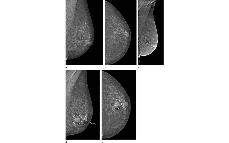
Images in a 72-year-old woman who was diagnosed with a 13-mm lymph node-negative invasive lobular carcinoma luminal B–like human epidermal growth factor receptor 2 breast cancer 18 months after a screening negative for cancer in the Malmö Breast Tomosynthesis Screening Trial. (a) Mediolateral oblique and (b) craniocaudal digital mammography (DM) images at screening. The slight retraction of the nipple was unchanged compared with previous DM screening images. c) Digital breast tomosynthesis at screening. DM images of (d) mediolateral oblique and (e) craniocaudal views at diagnosis, small marker at lump location. Increased nipple retraction (arrow) and central mass (circle on d and e).