New Consensus Statement Aims to Improve Endometriosis Evaluation
Recommendations focus on enhancing deep endometriosis detection

A new Society of Radiologists in Ultrasound (SRU) expert consensus statement to improve endometriosis evaluation was published in the journal Radiology.
The consensus provides recommendations for augmenting routine pelvic US through additional maneuvers and imaging to improve diagnosis of deep endometriosis.
Endometriosis is a prevalent and potentially debilitating condition estimated to affect 10% of women of reproductive age and occur in 21% of women undergoing hysterectomy with chronic pelvic pain. In the U.S., there is an over seven-year delay between the onset of symptoms and a diagnosis of endometriosis.
Endometriosis is also associated with infertility and subfertility, affecting 20-50% of patients with these conditions. Deep endometriosis, extending to any depth beneath the peritoneal surface, is the most severe form of endometriosis.
US is usually the first-line imaging modality used when patients report chronic pelvic pain or have issues of infertility, but few centers in the U.S. use it to screen for deep endometriosis. Existing scan protocol limitations and lack of awareness lead to suboptimal detection of deep endometriosis on pelvic US.
Scott W. Young, MD, and Rosanne M. Kho, MD, discuss the new expert consensus statement by the Society of Radiologists in Ultrasound to improve endometriosis evaluation.
SRU convened a multidisciplinary panel of experts to make recommendations aimed at improving the screening process for endometriosis.
“The purpose of this consensus panel is to recommend methods that increase the diagnostic sensitivity for endometriosis on pelvic ultrasound by increasing awareness, improving interpretation, adding simple techniques that are high yield for deep endometriosis, and improving protocols to triage patients,” said the statement’s first author Scott W. Young, MD, diagnostic radiology consultant, Division of Ultrasound, at the Mayo Clinic in Phoenix.
The panel was composed of experts in the imaging and management of endometriosis, including radiologists, sonographers, gynecologists, reproductive endocrinologists and minimally invasive gynecologic surgeons. A comprehensive literature review combined with a modified Delphi technique achieved a consensus.
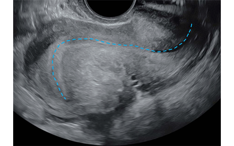
Transvaginal US in a 41-year-old patient with chronic pelvic pain and dyschezia shows the “question mark sign” uterine configuration. Longitudinal view shows an abnormal uterine configuration in which the uterus is sharply retroflexed because of deep endometriosis that is tethering the posterior cervix to the uterine corpus. This observation is usually identified by abnormal endometrial axis with sharp retroflection of the uterine fundus (dashed blue line) and constitutes a category B (ie, indirect endometriosis) observation.
https://doi.org/10.1148/radiol.232191 © RSNA 2024Guidelines Target Symptomatic Patients at Typical Risk
“The statement defines the targeted screening population, describes techniques for augmenting pelvic ultrasound, establishes direct and indirect observations for endometriosis on ultrasound, creates an observational grading and reporting system and makes recommendations for additional imaging and patient management,” Dr. Young said.
Panel recommendations include transvaginal US of the posterior compartment, observation of the relative positioning of the uterus and ovaries, and the uterine sliding sign maneuver to improve the detection of endometriosis.
“These additional techniques typically can be performed in less than five minutes and could ultimately decrease the delay of an endometriosis diagnosis in at-risk patients,” Dr. Young said.
The panel also recommends that direct and indirect observations of deep endometriosis should be assessed during the exam, and results should be reported using four categories: Incomplete (APU-0), Normal (APU-1), Equivocal (APU-2) and Positive (APU-3) with associated management recommendations.
“The SRU consensus on routine pelvic ultrasound for endometriosis aims to enhance deep endometriosis detection even at an initial ultrasound and with minimal additional time during imaging and no special patient preparation,” Dr. Young said. “Focusing imaging on anatomic regions where deep endometriosis is common can increase detection and decrease diagnostic delay.”
Dr. Young noted that these guidelines are meant for symptomatic patients at typical risk for endometriosis. Patients at high risk because of prior diagnostic or therapeutic laparoscopy for endometriosis or strong clinical indications may benefit from proceeding directly to advanced endometriosis imaging, particularly if they are likely to undergo surgery or if monitoring is needed in the setting of infertility and medical treatment.
The authors advise that validation studies will be necessary to prove the accuracy of augmented pelvic US in widespread clinical application.
For More Information
Access the Radiology article, “Society of Radiologists in Ultrasound Consensus on Routine Pelvic US for Endometriosis.”
Read previous RSNA News stories about US imaging: