4D Flow MR Comprehensive Mapping of Cerebral Hemodynamics by Age
Data can be used to understand how cardiovascular and dementia risk factors are related to normal vascular aging


Previous evidence has suggested that cerebral blood flow decreases with age, but a new study using 4D flow MR imaging bolsters these findings in a large cohort—and has allowed the investigators to break down age ranges as finely as five years.
“4D flow is a powerful and precise tool for measuring blood flow, and it’s regarded to be more accurate than measures based on transcranial Doppler or perfusion imaging. Yet there is still substantial variation in brain flow as a function of age and sex,” said coauthor Laura B. Eisenmenger, MD, associate chief of MRI and medical director of imaging services for the Wisconsin Institutes for Medical Research in Madison. “This study has really highlighted the need to understand this heterogeneity. Of course, using age as a continuous variable is often limited by sample sizes. Our cohort size enabled this analysis, showing that aging leads to reductions in blood flow and higher pulsatility indicating vessel stiffening.”
Strong Correlations with Age
For the study, published in Radiology, patients were selected from the Wisconsin Alzheimer Disease Research Center and Wisconsin Registry for Alzheimer Disease Prevention.
While the registry has enabled novel discoveries related to Alzheimer disease, the studies also included patients with normal cognitive findings, and this provided a large database to investigate flow changes in normal aging.
“We hope that our normative studies will establish quantitative baselines for clinical use of 4D flow,” she said.
Dr. Eisenmenger’s team examined cerebrovascular flow rates and pulsatility indexes in 759 middle-aged and older adults, mapping 17 major vessel segments for the group and further analyzing them by age and biologic sex.
Overall, the participants demonstrated a 4 mL/minute decrease in total cerebral blood flow for each year of age. The researchers noted no evidence of flow differences between sexes, but found that pulsatility tended to be higher in female patients.
“Perhaps the most important takeaway for practicing radiologists is that 4D flow is now a feasible imaging tool for evaluating intracranial hemodynamics,” Dr. Eisenmenger said.“This study has really highlighted the need to understand this heterogeneity. Of course, using age as a continuous variable is often limited by sample sizes. Our cohort size enabled this analysis, showing that aging leads to reductions in blood flow and higher pulsatility indicating vessel stiffening.”
– Laura B.Eisenmenger, MD
4D Flow MRI Beneficial To Aging Patients
The study stands out as a comprehensive survey of normative cerebrovascular flow rates and helps to establish baselines for normal flow according to age, commented University of Toronto professor of mechanical and industrial engineering David A. Steinman, PhD, in an accompanying Radiology editorial.
“Especially for the many researchers like myself who are seeking to deploy computer simulations to aid clinical decision-making, these tabulated data provide, for the first time, deep descriptive statistics that will allow us to confidently reflect—and communicate to patient and clinical stakeholders—the uncertainties of generic assumptions we are often forced to make about blood flow rates, which are still rarely acquired as part of clinical routine,” Dr. Steinman added.
In what was previously the largest study of this kind, a study in the Journal of Cerebral Blood Flow & Metabolism included 94 volunteers. The investigators relied on 2D cine phase-contrast MR and had to prescribe 13 individual arterial sections, requiring about an hour of scan time for each participant, noted Dr. Steinman.
With 4D flow, Dr. Eisenmenger’s team managed to shave the acquisition time to between 5.6 and 7.1 minutes for the entire head, plus an additional five minutes for manual vessel selection and automatic post-processing.
“Even in busy radiology workflows, the cost of those extra 10 minutes may come to be outweighed by the benefits of such rich data,” Dr. Steinman observed.
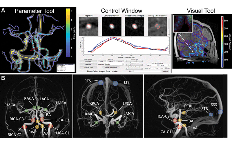
A contrast-unenhanced four-dimensional (4D) flow MRI data set in a 67-year-old female patient without cognitive impairment. (A) Semiautomated cranial 4D flow MRI postprocessing platform. The parameter tool (left) is an interactive three-dimensional interface with color-coded vessel centerlines overlayed on a semitransparent angiogram. Individual vessel segments can be selected for hemodynamic analysis with this tool. The control window (middle) shows local (in-plane) cross-sectional images of magnitude, velocity, complex difference, and segmentation contours, and flow profiles. This information is updated in real time when a vessel is selected from the parameter tool. Blood flow profiles over the cardiac cycle are displayed for the current centerline (black line) and neighboring centerlines (red and blue lines). The built-in visual tool (right) can display three-dimensional velocity vector arrows or “glyphs” within the segmented angiogram. Axial, sagittal, and coronal two-dimensional conventional magnitude sections, simultaneously obtained from the 4D flow MRI examination, are shown. Velocity arrows (image inset in visual tool) are color-coded by velocity magnitude and point in the direction of blood flow. (B) Four-dimensional flow MRI angiograms show vessel locations for hemodynamic measurements performed in this study. Coronal, axial, and sagittal maximum intensity projections (MIPs) from the three-dimensional complex difference data set, which is used for vessel segmentation during postprocessing, are shown. ACA = anterior cerebral artery, BA = basilar artery, ICA = internal carotid artery (C1 or C3 segment), L = left, MCA = middle cerebral artery, PCA = posterior cerebral artery, R = right, SSS = superior sagittal sinus, STR = straight sinus, TS = transverse sinus, VA = vertebral artery.
https://doi.org/10.1148/radiol.222685 © RSNA 2023
Vascular Aging Is Relevant to Cerebrovascular and Neurologic Health
The authors acknowledged limitations of their study, noting that they didn’t exclude patients with conditions such as hypertension and diabetes or a history of smoking. They also noted that the study sample leaned toward a higher risk of Alzheimer disease and acknowledged a lack of diversity in the sample as their patients were mostly female and white.
Additionally, the need to manually select vessel segments—though the postprocessing was mostly automated—might impede translation into clinical practice. Nevertheless, their findings mark a significant step toward establishing cerebral flow as a biomarker in normal aging.
In addition to highlighting the heterogeneity of cerebral flow dynamics with normal aging, Dr. Eisenmenger added that their work has inspired further studies at their institute to delineate fundamental variation from cardiovascular remodeling and the variations from technical factors such as image noise or a patient’s physiologic state.
While hemodynamic imaging using perfusion is often used to evaluate stroke and traumatic injuries, the measures are generally qualitative, continued Dr. Eisenmenger. Quantitative mapping could enable radiologists to diagnose vascular disease that is less obvious. It could also help to monitor flow changes following surgical and pharmaceutical interventions.
Ultimately, the authors speculated, large collections of correlated hemodynamic data and patient biometric data could be used to better understand how cardiovascular risk factors and dementia risk factors—such as AP0E4 mutations, which are associated with amyloid and tau production in the brain—are related to normal neurovascular aging.
“Future studies could apply methods such as ‘robust norms’ or Gaussian mixture modeling to detect vascular disease subpopulations, apply atlas-based or deep learning methods to completely automate postprocessing, and use temporal filtering to improve pulsatility index calculations,” they write.
“It will require substantial study to characterize variations across sites, individuals, diseases and populations,” Dr. Eisenmenger added.
Dr. Steinman agrees.
“Statistical modelling and deep learning are only ever as good as the data they’re being fed, and they have voracious appetites,” he concluded.
For More Information
Access the Radiology study, “Normative Cerebral Hemodynamics in Middle-aged and Older Adults Using 4D Flow MRI: Initial Analysis of Vascular Aging,” and the accompanying editorial, “Comprehensive Atlases of Intracranial Blood Flow Rates: A Hard Nut Finally Cracks?.”
Access the Journal of Cerebral Blood Flow & Metabolism.