Lung Damage May Persist Long After COVID-19 Pneumonia
Study underscores radiology's role in identifying at-risk patients and assisting in follow-up and management


Some people recovering from COVID-19 pneumonia have CT evidence of damage to their lungs that persists a full year after the onset of symptoms, according to a new study published in Radiology.
The COVID-19 pandemic has considerably increased the demand for acute and post-acute healthcare worldwide.
COVID-19’s short-term effects on the lungs, such as pneumonia, are well documented. Much less is known about the illness’ long-term effects on the lungs.
As part of an Austria-based observational study on the development of lung disease in patients with SARS-CoV-2 infection, researchers looked at patterns and rates of improvement of chest CT abnormalities in patients one year after COVID-19 pneumonia.
The researchers assessed lung abnormalities on chest CT in 91 participants, mean age 59 years, at several points over one year after the onset of COVID-19 symptoms.
At one year, CT abnormalities were present in 49, or 54%, of the 91 participants. Of these 49 participants, two (4%) had received outpatient treatment only, while 25 (51%) were treated on a general hospital ward and 22 (45%) had received intensive care unit (ICU) treatment.
“The observed chest CT abnormalities from our study are indicative of damaged lung tissue,” said study co-author Anna Luger, MD, from the Department of Radiology at Innsbruck Medical University in Innsbruck, Austria. “However, it is currently unclear if they represent persistent scarring, and whether they regress over time or lead to pulmonary fibrosis.”
While CT abnormalities decreased in initial follow-ups, 63% of participants with abnormalities did not show any further improvement after six months. Age over 60 years, critical COVID-19 severity and male gender were associated with persistent CT abnormalities at one year.
CT Shows Residual Chest Abnormalities
Evidence from the SARS-CoV-1 outbreak of 2002 to 2004 shows that lung abnormalities may remain detectable even after decades, but do not show any progression, according to study co-author Leonhard Gruber, MD, from the Department of Radiology at Innsbruck Medical University. Recent studies, though, have shown a risk of progression of lung abnormalities such as the ones depicted on CT.
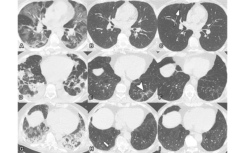
Serial non-contrast axial chest CTs of three study participants with prior COVID-19 pneumonia. Chest CT of a 44-year-old man (upper row, A-C) displayed extensive bilateral GGO and supleural reticulation during acute COVID-19 (A). At the 2-month follow-up almost complete resolution of GGO with residual subpleural reticulation in the middle lobe was noted (B). These subpleural reticulations (arrow) persisted up to one year after onset (C). Chest CT of a 68-year-old-man (middle row, D-F) demonstrated patchy bilateral consolidations, a subpleural arcade-like sign and pleural effusions during active infection (D). At the 2-month follow-up, a substantial improvement of OP pattern was noted with GGO and subpleural reticulation including arcade-like sign (arrowhead) in the left lower lobe (E). At the 1-year follow-up, further improvement was noticed. However, subtle reticulation and GGO could still be detected (F). Chest CT of a 79-year-old man (lower row, G-I) displayed bilateral consolidations and small areas of GGO while admitted to the ICU (G). At the 2-month follow-up, residual GGO and small subpleural microcystic changes (thick arrow) were noticed (H), which persisted up to 1 year after onset (I).
Luger et al, Radiology 2022; In Press, ©RSNA 2022
“In a recently published clinical study of our CovILD interdisciplinary working group, we were able to show that the severity of acute COVID-19, protracted systemic inflammation and the presence of residual chest CT abnormalities are strongly related to persistently impaired lung function and clinical symptoms,” said study co-author Christoph Schwabl, MD, from Innsbruck Medical University.
The study underscores radiology’s role in helping identify patients at risk for post-COVID-19 consequences and assisting in COVID-19 follow-up management.
“In the end, long-term follow-up, both clinical and radiological, is necessary to gather more information about the course and clinical role of persisting SARS-CoV-2 related chest CT abnormalities,” said study senior author Gerlig Widmann, MD, chief thoracic radiologist at Innsbruck Medical University.
The researchers intend to continue gathering data on patients with persistent CT abnormalities.
For More Information
Access the Radiology study, “Chest CT of Lung Injury 1 Year after COVID-19 Pneumonia: The CovILD Study.”
Access the related Radiology editorial, “COVID-19 Pandemic: The Road to Recovery.”
Read previous RSNA News articles related to COVID-19: