COVID-19 Linked to Potentially Dangerous Eye Abnormalities
Study Demonstrates Need for Eye Screening for COVID-19 Patients

Researchers using MRI have found significant abnormalities in the eyes of some people with severe COVID-19, according to a study in Radiology.
The COVID-19 pandemic has affected more than 100 million people since it began early in 2020. While the virus primarily attacks the lungs, it has been linked with eye abnormalities like conjunctivitis and retinopathy. Eye abnormalities visible on MRI exams have been reported but there is limited research on the nature and frequency of these abnormalities.
Eye Screening Can Lead to Earlier Treatment for COVID-19 Nodules
To find out more, the French Society of Neuroradiology (SFNR) initiated a study of 129 patients with severe COVID-19 who underwent brain MRI.
Of the 129 patients, nine (7%) had abnormal MRI findings of the globe, or eyeball. The MRI scans showed one or more nodules in the back part, or posterior pole, of the eyeball. Eight of the nine patients had spent time in the intensive care unit (ICU) for COVID-19.
“We showed that a few patients with severe COVID-19 from the French COVID-19 cohort had one or several nodules of the posterior pole of the globe,” said study lead author Augustin Lecler, MD, PhD, associate professor at the University of Paris and neuroradiologist from the Department of Neuroradiology at the Foundation Adolphe de Rothschild Hospital in Paris. “This is the first time these findings have been described using MRI.”
All nine patients had nodules in the macular region and eight had nodules in both eyes.
The results suggest that screening should be considered in all patients with severe COVID-19 to detect these nodules. In clinical practice, this screening could include dedicated exploration of the eyes with high-resolution MRI, the researchers said. Additional recommended exams include fundoscopy, which uses a magnifying lens and a light to check the back of the inside of the eye, and optical coherence tomography, a noninvasive test that provides a 3D picture of the structure of the eye.
Dr. Lecler noted that severe eye problems might largely go unnoticed in the clinic, as COVID-19 patients hospitalized in the ICU are often being treated for much more severe, life-threatening conditions.
“Our study advocates for screening of all patients hospitalized in the ICU for severe COVID-19,” Dr. Lecler said. “We believe those patients should receive specific eye-protective treatments.”
The mechanism behind nodule formation remains unknown, the researchers said, although it could be related to inflammation triggered by the virus. Inadequate drainage of the veins of the eyes, a problem found in patients who spend time in the ICU in the prone position or intubated, may also be a factor. Seven of the nine patients with eye abnormalities in the study had been placed in a prone position in the ICU for an extended time.
The researchers are performing follow-up clinical and MRI examinations in the survivors to monitor the nodules and see if they carry any clinical consequences such as vision loss or visual field impairment.
They are also performing MRI examinations in new patients with severe COVID-19 from the second and third waves of the pandemic, using more comprehensive ophthalmological tests to correlate with the MRI results.
The effects on patients with moderate COVID-19 are currently under investigation.
“We have launched a prospective study with dedicated high-resolution MR images for exploring the eye and orbit in patients with light to moderate COVID,” Dr. Lecler said. “Therefore, we will be able to know whether our findings were specific to severe COVID patients or not.”
The findings support previous research that showed COVID-19 exacts a greater toll in people with existing health problems. Of the nine patients with eye nodules, two had diabetes, six were obese and two had hypertension.
For More Information
Access the Radiology study, “Ocular MRI Findings in Patients with Severe COVID-19: A Retrospective Multicenter Observational Study.”
Read previous RSNA News stories on COVID-19:
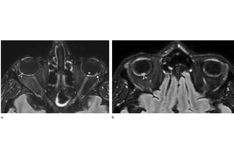
56-year-old man presenting with severe COVID-19. Diagnosis of SARS-CoV-2 infection was based on a positive quantitative real-time RT-PCR test for SARS-CoV-2 nucleic acid performed on nasopharyngeal and lower respiratory tract swabs. The patient had been hospitalized in intensive care unit for 20 days when an MRI was performed due to delayed awakening despite discontinuation of sedation. He presented with acute respiratory distress syndrome, with a median Simplified Acute Physiology Score (SAPS II) of 45. He was intubated on high-flow supplementary oxygen and placed in the prone position. A, B, 3D FLAIR-weighted MRI reformatted in the axial plane showing several hyperintense nodules of the posterior pole of the globe located in the macular region (white arrowhead) and the extramacular region (black arrowheads). Note the presence of a focal temporal retinal detachment of the left eye (arrow).