Next-Generation MRI Moves to the Forefront as a Diagnostic Tool
Advancing technology brings benefits of MRI to growing number of patients

MRI is a powerful, continually advancing tool with quantifiable results that can benefit patients as a front-line choice for screening and diagnosis, researchers say.
“MR absolutely is not a stagnant field,” said Stephen J. Riederer, PhD, a medical physicist and professor of radiology at Mayo Clinic College of Medicine and Science in Rochester, MN and head of the MRI Laboratory at Mayo. “MR is still a tremendously exciting area to work in and it’s important for us to ask how we can make it even better, expand on it and explore specific areas where special-purpose machines might be useful.”
The Promise of 4D Flow MRI
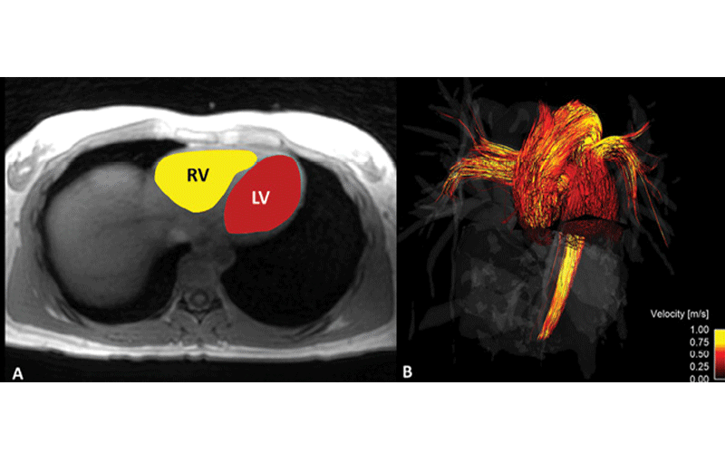
Advances in a powerful new technology, 4D flow MRI, offer a comprehensive picture of the heart and aorta. The fourth dimension, movement, allows for better visualization of blood flow through the cardiovascular system, potentially identifying areas that require follow up. A recent study published in Radiology: Cardiothoracic Imaging, “Two-Minute k-Space and Time–accelerated Aortic Four-dimensional Flow MRI: Dual-Center Study of Feasibility and Impact on Velocity and Wall Shear Stress Quantification,” demonstrated that 4D flow MRI can depict significant differences in how the heart contracts between men and women. (See image below.)
The study showed that kinetic energy during contraction was significantly higher in men, while vorticity and strain were higher in women. Because these results were found in healthy volunteers, the research team, led by Emilie Bollache, PhD, of the Department of Radiology, Northwestern University in Chicago proposed that blood flow metrics could be useful reference standards for evaluating disease.

Quantitative MR Parameters
Quantitative MR parameters have a foreseeable clinical impact for patients, as demonstrated by a February 2020 study, “Comparison of Abbreviated Breast MRI vs. Digital Breast Tomosynthesis for Breast Cancer Detection Among Women With Dense Breasts Undergoing Screening,” in the Journal of the American Medical Association, led by Christopher E. Comstock, MD, an attending radiologist and director of breast imaging clinical trials at Memorial Sloan-Kettering Cancer Center. The study explored ways to make breast MR screening more accessible to patients.
“There’s a movement toward vascular-based screening that’s based on tumor angiogenesis that, unlike mammography (based on structural imaging), is not limited by breast density,” Dr. Comstock said. “The problem in the past has been that even though we’ve known that MR is the most powerful tool, factors like the time in the scanner and cost has been limiting, so we would typically only offer the modality to high-risk gene carriers.”
In this trial, including more than 1,400 women, the aim was to compare invasive cancer detection rate of abbreviated breast MRI compared with DBT in women with dense breasts and to determine whether MR could be expanded as a screening tool to benefit more women.
“Previous research has involved doing a full MR scan, stripping out some of the sequences to turn it into an abbreviated study, and having readers examine it — so they were reader studies,” Dr. Comstock said. “But we examined how it performs in the average user’s hand using only abbreviated MR from the start.”
The research included approximately half academic and half community or private practice departments, exploring whether MR can be done in a short amount of time. The answer was yes, in about eight minutes. And the women tolerated it well; about 96% were able to complete the study. And reducing the acquisition and reading times drives a reduction of the cost of performing MR, which moves the modality closer to becoming a new paradigm in screening.
“Initially you might include ultrafast imaging, diffusion-weighted imaging and additional sequences for kinetics to improve MR specificity, but in subsequent screening years, we could just do a three-minute MR scan including only pre- and post-contrast sequences,” Dr. Comstock said. “There’s also the potential to perform non-contrast scans, which don’t even require an injection.”

Underdiagnosis is still a big problem in breast screening programs, Dr. Comstock said, with more than 40,000 women dying of breast cancer each year. “Screening has changed because of breast density legislation where a significant number of women now get mammography plus ultrasound, which is not very efficient,” Dr. Comstock said. “Despite screening with mammography and even mammography plus US, cancer is still missed.”
Drs. Comstock and Riederer both identified artificial intelligence (AI) as an area that needs more attention on the MRI front.
“There’s a lot of publicity about AI, and this is an area where not all the protocols are available, so improving ease of usage can make it more clinically applicable,” Dr. Riederer said. “But with MR there is already a lot of additional intelligence under the hood. You can implement intelligent calibration, intelligent selection of the receiver coils you use — and there are functions like patient-specific parameter selection, automated angulation and alternative reconstruction methods to make better use of the data.”
To make the most of MR in a clinical setting, Dr. Riederer cited examples where imaging centers can implement specialized applications.
“Typically, in hospitals, there’s a division between neuro and body MR, but even in those cases, machines can be devoted to musculoskeletal or abdominal imaging,” he said. “We also have moderate-bore 3T head scanners to accommodate imaging of the brain; the smaller bore makes siting easier and reduces helium usage; and the higher performance gradients enable faster scans and image quality improvement such as better diffusion imaging of the brain. There’s also interest in very low-field scanners, or ‘portable’ MRI. This is an area where the technology is enabling special echelon applications.”
“At the turn of the Y2K century, internists were asked to rank recent advances in medicine that were the most pivotal, and CT and MR were at the top of the list,” Dr. Riederer said. “We’re now looking at doing MR at 7 Tesla. We need to adopt the mindset that MR, where applicable and appropriate, doesn’t have to be the modality that is used second or third in the workup of a patient. We need to think of this as a front-line diagnostic tool.”
For More Information
Access the Radiology: Cardiothoracic Imaging study, “Two-Minute k-Space and Time–accelerated Aortic Four-dimensional Flow MRI: Dual-Center Study of Feasibility and Impact on Velocity and Wall Shear Stress Quantification.”
Access the JAMA study, “Comparison of Abbreviated Breast MRI vs Digital Breast Tomosynthesis for Breast Cancer Detection Among Women With Dense Breasts Undergoing Screening,” at JAMAnetwork.com.