Accelerated 4D MR Aortic Imaging Sequence Cuts Scan Time to Two Minutes
Radiology: Cardiothoracic Imaging study conducted at centers in London, Chicago


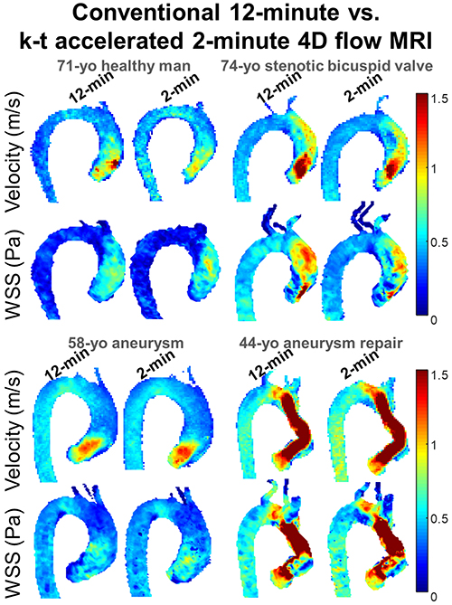
A four-dimensional (4D) k-space and time–accelerated MR imaging sequence shows promise for quantifying aortic velocity, with a scan time of only two minutes.
The study, recently published online in Radiology: Cardiothoracic Imaging, was an international project comprising patient cohorts and two centers, said lead author Emilie Bollache, PhD, a research associate at Laboratoire d’Imagerie Biomédicale at Sorbonne Université in Paris, who completed his post-doctoral fellowship at Northwestern University, Chicago.
“We developed this MR imaging sequence at Northwestern, which we shared with another center in London,” Dr. Bollache said. “It was exciting to assess the feasibility of this sequence at another center, and to see that it was really easy for them to use. We basically sent them the protocol and they installed the sequence on their own MR scanner, and it ran smoothly, and they could include patients very quickly.”
The study included 68 participants, either healthy volunteers or patients with aortic or cardiac disease. The participants underwent 4D imaging at a k-space and time at accelerated factor of 5. These scans were compared with conventional 4D flow MR scans — at an acceleration factor of 2— in patients within the same cohort.
Accelerated Sequence Proves Successful
The technique proved effective for depicting consistent healthy aging- and disease-related changes in aortic hemodynamics, Dr. Bollache said. The accelerated sequence was successfully reproduced at the second institution, London’s Barts Heart Centre.
“The 4D MR sequence is very powerful and can provide 3D visualization and quantification of blood flow through the heart and arteries,” Dr. Bollache said. “But the issue right now is that the scan time is too long. This new flow sequence is only two minutes long and it doesn’t require any navigator placement, so it’s also easy for technologists.”
The researchers found good agreement between the conventional and accelerated techniques, identifying significantly increased peak velocities and wall shear stress (WSS) in patients with stenotic or bicuspid valves compared with those in healthy volunteers. The team noted, however, that the accelerated technique significantly underestimated WSS and demonstrated significant differences in pulse wave velocity compared with the conventional technique.
“For more advanced parameters such WSS and pulse wave velocity, there needs to be more technical development to address the variance,” said study author Michael Markl, PhD, a professor and vice chair for research at the Department of Radiology at Northwestern University.
The authors speculated that the discrepancies could be explained by several factors, including respiratory motion and differences in spatial and temporal resolutions that could underestimate complex hemodynamic alterations. The researchers plan to investigate these factors.
“But for standard clinically used flow parameters, the accelerated flow sequence is ready for prime time in a true multi-center study,” Dr. Markl said.
Multi-Institutional Study Planned
The next step will be a study across 6 to 10 institutions, where the researchers will again develop a joint protocol that they will distribute among the participating centers, said Dr. Markl. “Then we can do a core lab-based analysis to show how it works across different institutions and different patient populations.” The authors further recommended that the preliminary multicenter effort should involve institutions both with and without experience in cardiovascular MR imaging, to confirm the feasibility of the sequence.
“4D flow MR is a complex imaging technique and it’s currently being relegated, to some extent, to the realm of research, simply because it takes a long time to collect the data,” Dr. Markl said. “With this 2-minute scan, it’s been simplified and made much more efficient so that there’s a chance to bring it to a clinical setting on a more routine basis.”
For Your Information
Access the study, “Two-Minute k-Space and Time–accelerated Aortic Four-dimensional Flow MRI: Dual-Center Study of Feasibility and Impact on Velocity and Wall Shear Stress Quantification.”
4D Flow Imaging Focus of Hot Topic Session at RSNA 2019
The Hot Topic Session, “4D Flow Imaging in Congenital and Acquired Cardiovascular Disease-Clinical Impact,” will be held on Thursday, Dec. 5, at RSNA 2019. Presentations focus on congenital heart disease, structural heart disease and aorta, followed by a panel discussion.
To register for this and other RSNA 2019 courses, go to Meeting.org.