RSNA Publishes Expert Consensus on COVID-19 Reporting
Statement to help radiologists recognize COVID-19 pneumonia and provide guidance on standardized language for CT findings

RSNA has published an expert consensus statement on reporting chest CT findings related to COVID-19. The statement, published in Radiology: Cardiothoracic Imaging, is endorsed by the RSNA, the American College of Radiology and the Society of Thoracic Radiology.
“We believe it is important to provide radiologists and referring providers guidance and confidence in reporting these findings and a more consistent framework to improve clarity,” said Suhny Abbara MD, editor of Radiology: Cardiothoracic Imaging and professor of radiology and chief of the Cardiothoracic Imaging Division at UT Southwestern Medical Center in Dallas, Texas. “Clear evidence-based communication among health care providers, including radiologists, is imperative to improving patient care during this pandemic.”
Statement To Serve As A Guide for Institution Discussions on COVID-19
Coronavirus disease 2019 (COVID-19), caused by severe acute respiratory syndrome coronavirus 2 (SARS-CoV-2), has become increasingly prevalent worldwide, reaching a pandemic stage in March 2020.
While routine screening CT for the identification of COVID-19 pneumonia is currently not recommended by most radiology societies, the number of CTs performed in patients under investigation for COVID-19 has increased. Some patients may have incidentally detected findings that could be attributable to COVID-19 pneumonia, requiring radiologists to decide whether to mention COVID-19 specifically as a differential diagnostic possibility.
COVID-19 pneumonia, which has a high mortality rate among the elderly and those with diabetes, hypertension, and other comorbidities, is spreading rapidly in communities. As a result, including “COVID-19” frequently in radiology report could trigger a cascade of events including infection control measures and anxiety for both the clinician and patient. Importantly, CT imaging features of COVID-19 can overlap significantly with other causes of acute lung injury and pneumonia, complicating interpretations.
The consensus statement, developed by experts at nine U.S. academic medical centers, aims to help radiologists recognize findings of COVID-19 pneumonia and provide guidance on reporting CT findings potentially associated with COVID-19, including standardized language to reduce reporting variability.
Standardized reporting language will improve communication with referring providers and has the potential to enhance efficiency and reduce anxiety in management of patients during the pandemic.
The paper discusses the potential role of CT in COVID-19, parameters for structured reporting, and the pros, cons and limitations of adopting this strategy.
Because practice patterns may vary by institution, the document is meant to serve as a guide. The authors recommend that radiologists consult with clinical colleagues at their institutions to establish a consensus reporting approach.
For More Information
Access the Radiology: Cardiothoracic Imaging statement, “Radiological Society of North America Expert Consensus Statement on Reporting Chest CT Findings Related to COVID-19. Endorsed by the Society of Thoracic Radiology, the American College of Radiology and RSNA.”
Access the RSNA COVID-19 Resources webpage. Visit and bookmark this page to access the latest guidance, original research, image collection and resources on COVID-19.
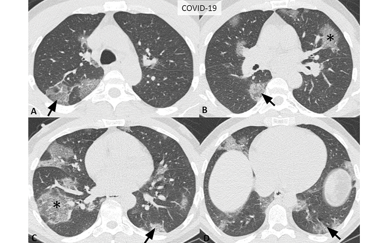
Typical CT imaging features for COVID-19. Unenhanced, thin-section axial images of the lungs in a 52-year-old man with a positive RT-PCR (A-D) show bilateral, multifocal rounded (asterisks) and peripheral GGO (arrows) with superimposed interlobular septal thickening and visible intralobular lines (“crazy-paving”).
Routine screening CT for diagnosis or exclusion of COVID-19 is currently not recommended by most professional organizations or the U.S. Centers for Disease Control and Prevention.