The Future of Thoracic Radiology

Soon after Roentgen’s discovery of the X-ray, chest fluoroscopy and chest radiography became an established clinical procedure. The standard chest radiograph remained the mainstay of chest imaging for almost 80 years until the advent of CT and MRI.
As Dr. Marilyn Siegel writes in a previous article regarding the future of pediatric radiology, “advances in technology have revolutionized imaging impacting the diagnosis and treatment of disease.” We can expect future changes in imaging resulting not only in qualitative improvement of images but also in the ability to provide both functional data and quantitative assessment. Such advances may allow more accurate diagnosis and better treatment and will add value to radiologic practice. This article provides a review of some recent advances in thoracic imaging technology, and discusses new methodologies that we may expect in thoracic radiology over the next decades.
Recent Advances in Imaging: Thoracic Imaging Technologies
Computed Tomography
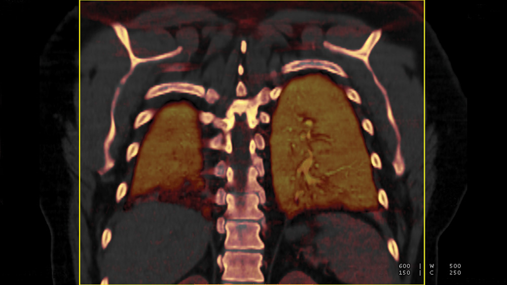
Over the past 30 years we have witnessed revolutionary advances in CT. These have included helical acquisition, reduced gantry rotation times, multidetector systems with submillimeter spatial resolution, advanced computational ability for image construction and the development of low dose scanning. Although CT has impacted thoracic radiology over the entire spectrum of thoracic disease, it has had the most impact in the areas of emphysema, interstitial lung disease, lung cancer and the diagnosis of pulmonary embolism.
The advent of CT pulmonary angiography provided an accurate tool to establish or rule out the diagnosis of pulmonary embolism and also has provided information about patient prognosis and clinical outcome.
One of the relatively recent and new challenges in thoracic imaging is in the area of lung cancer screening. Following the results of the National Lung Screening Trial (NLST), CT has been widely implemented nationwide for the screening of high risk patients for early lung cancer. Computer-aided diagnosis (CAD) has become an important tool in nodule detection, characterization and evolution. The implementation of CAD systems had been somewhat limited because of difficulties in integrating work flow with current PAC systems. However, computer-aided detection and diagnosis will be an important implementation tool for future utilization.
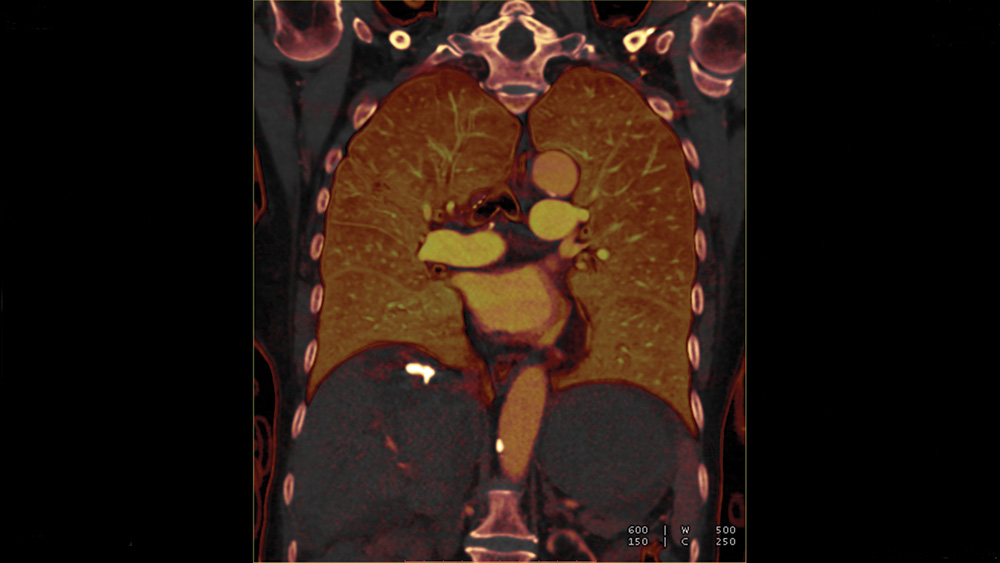
One of the most important recent advances in thoracic radiology and in vascular imaging in general has been the implementation of dual-energy CT.
Dual-energy CT allows the differentiation of materials with large atomic numbers such as iodine. Dual energy CT angiography can evaluate perfusion defects in cases of pulmonary embolism.
Although not as sensitive and specific as scintigraphy, it is highly accurate when there is complete occlusion of pulmonary arteries. The iodine perfusion map may also be used for the follow-up of pulmonary embolism. Dual energy CT may also be helpful in the evaluation of pulmonary nodules.
Using dual-energy CT, solitary pulmonary nodules can be characterized and malignant pulmonary nodules may be distinguished from benign lesions by providing information on the degree of enhancement and the presence of calcification.
MRI
The thorax has been one of the most challenging applications for the use of MRI, and it has been traditionally applied to the assessment of mediastinal and pleural diseases. Recent technical advances in sequencing, scanners and coils, adaptation of parallel imaging techniques, utilization of contrast media, and the development of post processing tools has enhanced the applications of MRI in the thorax.
Thoracic MR can now be substituted for traditional imaging techniques or can play a complimentary role in the management of patients with various chest diseases.
Continuous motion from cardiac vascular pulsation and respiratory motion have been the major challenges for MRI of the chest. More recently, MRI has evolved from a research tool to an effective modality in the assessment of lung disease. Technical advances such as very short echo times and ultrafast turbo spin echo acquisitions that allow breath holding imaging with full anatomic coverage have improved the capability of thoracic MRI. The use of contrast agents for perfusion MRI and gas imaging for assessment of pulmonary ventilation have further increased the applications of MRI in the investigation of lung diseases.
Other important applications include the detection of pulmonary nodules and the management of patients with various thoracic malignancies. New MR techniques and sequences as well as diffusion weighted images and dynamic contrast enhanced techniques have been shown to be helpful in nodule evaluation and lymph node staging in lung cancer.
PET/CT
Advances in FDG-PET/CT have had an increasing role in nonmalignant chest diseases such as active granulomatous infections and inflammation. Inflammatory conditions are frequently confused with malignancy. However PET can be very helpful in providing suggestive patterns of activity with certain inflammatory diseases such as IgG4-related diseases and vascular disease.
Also on the horizon is the development of new approved radiotracers. Not all are applicable to the thorax, but new octreotide related PET scan agents, which have been well studied in Europe, and now are available in the U.S., may allow for better clinical management of several thoracic diseases including neuroendocrine tumors such as carcinoid tumors. Major advances in PET imaging technology will involve new detector systems and new post processing algorithms.
Although PET/MR has arrived it is mainly a research tool at the present time. Its applications in the thorax remain to be explored.
THERESA C. McLOUD, MD, served as the divison head of thoracic radiology from 1982 to 2001 and is currently vice chair for education in the Department of Radiology and the incumbent Manorama and Virender Saini Endowed Chair in Radiology Education, at Massachusetts General Hospital (MGH), Boston.
A Boston native, Dr. McLoud earned her medical degree from the McGill University Faculty of Medicine in Montreal, completed a thoracic imaging fellowship at the Yale University School of Medicine and became an assistant professor of diagnostic radiology at Yale. In 1976, she returned to Boston and joined Harvard Medical School where she has been professor of radiology since 1993. Dr. McLoud is the first woman in the MGH Department of Radiology to hold the rank of professor at Harvard.
A world-renowned expert in thoracic imaging, Dr. McLoud has conducted more than 150 postgraduate courses and published more than 200 scientific papers, reviews, and book chapters. Among her many awards, Dr. McLoud has received the Association of Program Directors in Radiology Achievement Award and the Marie Curie Award from the American Association for Women Radiologists. Dr. McLoud served as the 2008 RSNA President and received the RSNA Gold Medal in 2013.