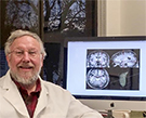
MRI Shows Early Brain Changes in Autistic Children

While researchers continue to make progress in understanding autism spectrum disorder (ASD), the statistics on ASD remain sobering.
In the U.S., approximately one in 68 children has been identified with some form of ASD, according to the Centers for Disease Control and Prevention. The risk is even higher — as much as one in five — for infants with an autistic older sibling.
Early diagnosis and intervention, which can improve an ASD patient’s symptoms and ability to function, is critical. As recent research demonstrates, imaging plays an increasingly pivotal role in early diagnosis and intervention in ASD.
Researchers have demonstrated that MRI shows changes in the brain related to autism as early as six months into a child’s life, potentially speeding interventions that might reduce the severity of the disorder in some patients. The findings were published in the journal Nature.
“Autism is a life-long condition, and if we are able to intervene early, children with autism do much better,” said study co-author Stephen R. Dager, MD, a radiology professor at the University of Washington in Seattle. “Using a combination of imaging and behavioral approaches to identify children who are at risk of autism could have a huge impact on their lives and the lives of the people around them.”
Experts agree that the findings show the potential for brain MRI in children facing a high risk of ASD, a complex developmental disorder that affects a person’s ability to communicate and interact with others.
“Having a biomarker for early identification of ASD will expand the window of opportunity for targeted intervention during critical periods of brain development and may ultimately improve outcomes for these children,” said Christopher T. Whitlow, MD, PhD, MHA, a member of the RSNA Public Information Advisors Network (PIAN), and an associate professor of radiology at Wake Forest School of Medicine and a radiologist at Wake Forest Baptist Medical Center in Winston-Salem, N.C.
Researchers at four different clinical sites across the U.S., including the University of Washington, used MRI to build on previous findings of early cerebral enlargement in children with ASD.
The study comprised 106 infants at high familial risk of ASD and 42 low-risk infants. Researchers used the same MRI scanner platform, operating system, hardware and head coils across all four sites and deployed a phantom to obtain structural dimensions and calibrate the scanner.
The results showed that 15 high-risk infants who were diagnosed with autism at 24 months experienced a hyper-expansion of brain surface area from 6 to 12 months, as compared with high-risk babies who did not show evidence of autism at 24 months.
This spurt in brain growth was linked to the emergence and severity of autistic social deficits.
“The study suggests that even as early as six months, we’re able to identify subtle behavioral and imaging differences that retrospectively identify those children with a diagnosis of autism at age two,” Dr Dager says.
Algorithm Advances ASD Research
Researchers used MRI data to develop a deep-learning algorithm that classified babies most likely to meet ASD criteria at age 24 months based primarily on surface area information from earlier brain MRIs. When they applied the algorithm to a separate set of babies, they successfully identified 80 percent of those infants who would be clinically diagnosed with autism at age 24 months.
This information would be helpful to parents who already have an autistic child who want to know their new baby’s risk of developing ASD as soon as possible. Babies at highest risk could begin interventions earlier, before ASD is present and when the brain is most malleable. Such interventions may have a greater chance of improving outcomes than treatments started after diagnosis.
While further research is necessary, Dr. Whitlow says that this study is important for a number of reasons.
“It shows the power of combining quantitative structural MRI measures and deep learning algorithms to identify patterns of brain development that might otherwise be difficult to discern using conventional qualitative assessment,” Dr. Whitlow said.
He added that targeting relatively conventional structural MRI measures for this study may facilitate rapid translation of these findings into clinical practice.
Web Extras
For more information on the study, "Early Brain Development in Infants at High Risk for Autism Spectrum Disorder," go to http://www.nature.com/nature/journal/v542/n7641/full/nature21369.html