Brain Changes Seen in Youth Football Players without Concussion
An increase in subconcussive head impact exposure may have an effect on white matter (WM) integrity in youth athletes, even in the absence of a clinically diagnosed concussion, according to new Radiology research.
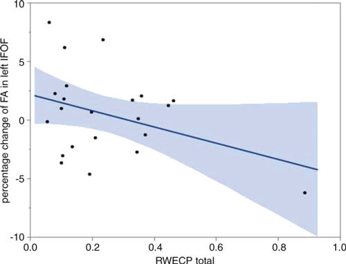
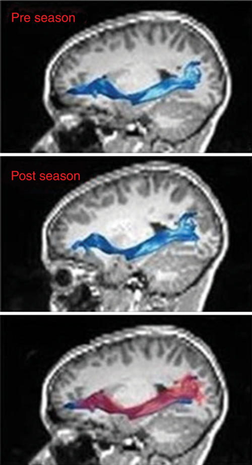
Naeim Bahrami, PhD, from the Advanced Neuroscience Imaging Research (ANSIR) Laboratory in Winston-Salem, N.C., and colleagues found a statistically significant relationship between combined-probability risk-weighted cumulative exposure (RWECP) and change of fractional anisotropy (FA) in the left inferior fronto-occipital fasciculus (IFOF).
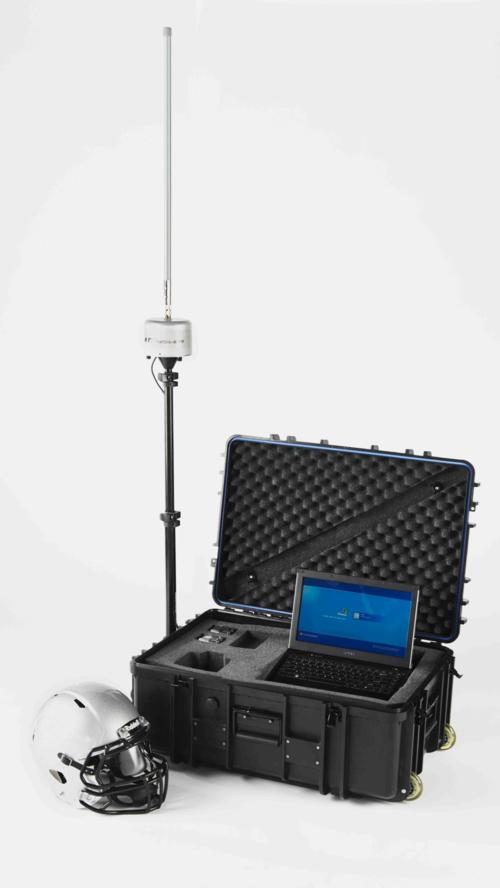
In a study of 25 male participants (age range 8 to 13 years) from a local youth football league, researchers used the Head Impact Telemetry system to record head impact data and quantify the RWECP There were statistically significant linear relationships between RWECP and decreased FA in the whole, core and terminals of left IFOF. A trend toward statistical significance in right superior longitudinal fasciculus (SLF) was observed. A statistically significant correlation between decrease in FA of the right SLF terminal and RWECP was also observed.
“The results of this study suggest that subconcussive impacts can result in changes in the WM microstructure of the IFOF and SLF fiber bundles,” the authors write.


