Heart Disease Protein Linked to Brain Damage
In community-dwelling middle-aged and elderly persons, subclinical cardiac dysfunction as reflected by serum N-terminal pro–B-type natriuretic peptide (NT-proBNP) levels is associated with global and microstructural MRI markers of subclinical brain damage, new Radiology research shows.
Hazel I. Zonneveld, MD, of Erasmus MC University Medical Center in Rotterdam, the Netherlands, and colleagues measured serum levels of NT-proBNP in 2,397 participants without dementia or stroke and without clinical diagnosis of heart disease who were drawn from the population-based Rotterdam Study.
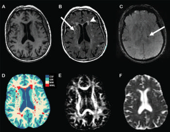
They found a higher NT-proBNP level was associated with smaller total brain volume and was predominantly driven by gray matter volume. Higher NT-proBNP level was associated with larger white matter lesion volume, with lower fractional anisotropy and higher mean diffusivity of normal-appearing white matter.
“Our findings suggest that the heart and brain are intimately linked, even in presumably healthy individuals. This is essential since cardiac dysfunction and subclinical brain damage are growing problems,” the authors write.

Web Extras
- Access the study, "N-Terminal Pro–B-Type Natriuretic Peptide and Subclinical Brain Damage in the General Population," at http://pubs.rsna.org/doi/full/10.1148/radiol.2016160548