Multiphase CT Shows Promise for Stroke Patients
Multiphase CT is a faster, simpler tool for treating stroke patients, saving critical time and possibly lives, new research shows


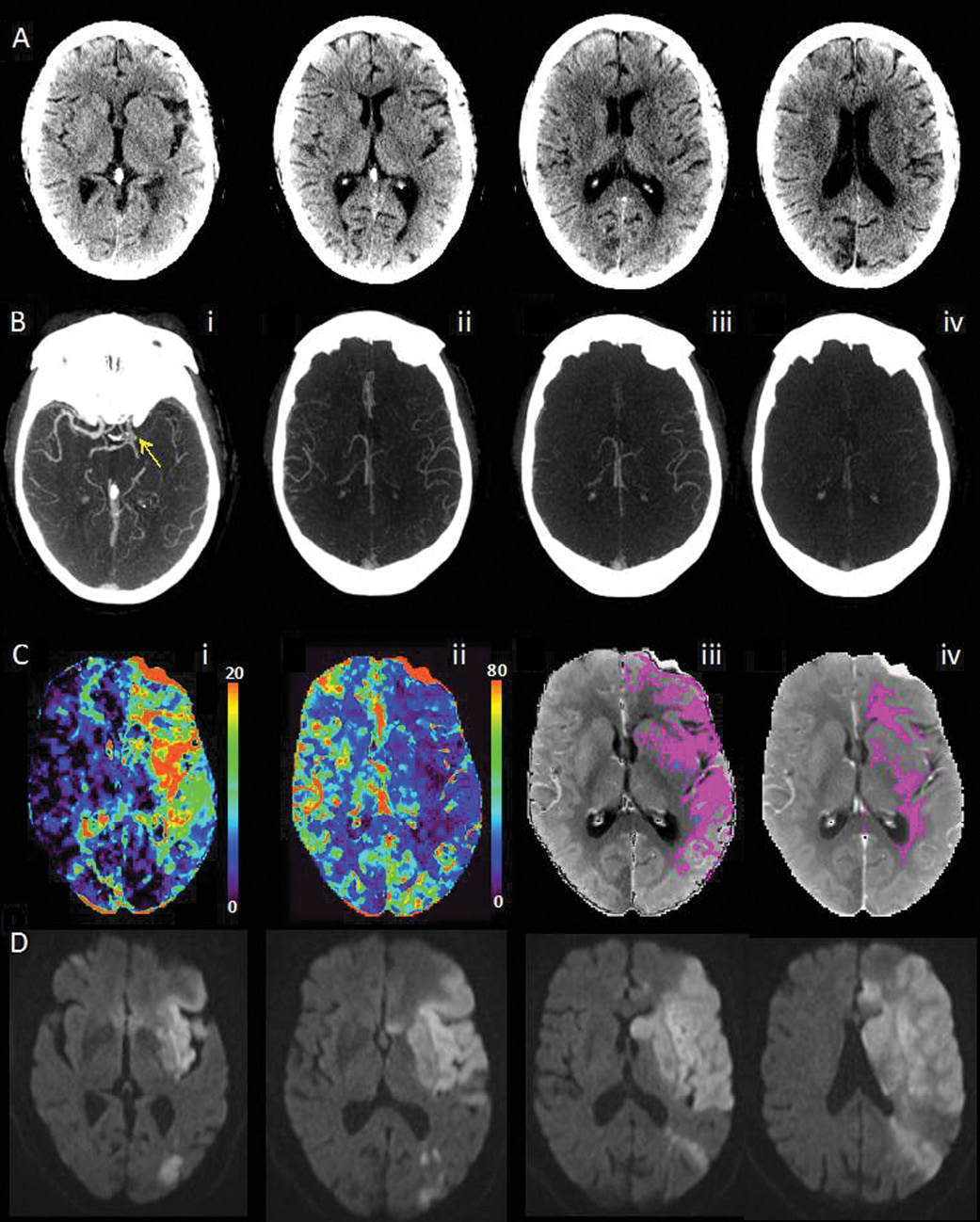
A new brain imaging technique—multiphase CT angiography—allows physicians treating acute ischemic stroke (AIS) patients to make faster decisions at a time when every second is critical, new Radiology research shows.
The imaging tool quickly produces crucial data on collaterals, reducing uncertainty in clinical decision-making and allowing for slightly better prediction of clinical outcome than current imaging techniques, researchers say. The study appears in the May 2015 issue of Radiology.
“Multiphase CT angiography allows for consistency across different machines and different locations and leads to a quick diagnosis in terms answering the question, ‘Is this patient a suitable candidate to take to the next level and try for early recanalization?’” said author Mayank Goyal, M.D., director of research in the Department of Diagnostic Imaging at the University of Calgary in Canada.
While current techniques have allowed for significant progress in stroke treatment, they also possess disadvantages: conventional angiography is invasive and time-intensive, single-phase CT angiography lacks temporal resolution, perfusion CT and MRI are susceptible to patient motion and require trained personnel to process data, and dynamic CT angiography requires post-processing and whole-brain perfusion CT.
Multiphase CT angiography provides information on degree and extent of pial arterial filling in the whole brain more quickly than other methods—critical considering that every 30-minute delay in treatment could increase the risk of poor clinical outcome by approximately 14 percent, researchers said.
“This tool is easy to use and very fast to interpret,” said lead author Bijoy K. Menon, M.D., an assistant professor of neurology at the University of Calgary. “It can save critical time that can mean the difference between walking and not walking, talking or not talking, or even living or not living.”
Quick to Perform, Easy to Interpret
Researchers studied 147 acute ischemic stroke patients—all of whom underwent baseline unenhanced CT, single-phase CT angiography of the head and neck, perfusion CT and multiphase angiography CT, and compared techniques with respect to their interrater reliability and predictive ability. The mean patient age was 72 years and the patient cohort was about 50 percent male.
The authors independently assessed multiphase CT angiography in 30 randomly chosen subjects by using a six-point ordinal scale that was then reclassified into three clinically relevant categories ("good," "intermediate" and "poor" pial arterial filling). Researchers made treatment recommendations based on images using all scanning techniques.
Results showed that for intravenous tissue plasminogen activator decision making, there was agreement 92.5 percent (k = 0.68) of the time between single-phase vs. multiphase CT angiography—the highest rate of agreement. The next highest agreement rate was between unenhanced CT and multiphase CT angiography at 89.1 percent (k = 0.4). Similarly for intra-arterial treatment decisions, maximal agreement (89.8 percent, k = 0.8) was seen between single- and multiphase CT angiography.
Additionally, multiphase CT models decreased National Institutes of Health Stroke Scores by 50 percent over a 24-hour period.
Results showed that multiphase CT angiography has the ability to predict stroke outcomes slightly better than models using single-phase CT angiography and perfusion CT. In addition, the technique was quick to perform and produced images that were easy to acquire and interpret.
“It’s those in-between situations where we need to have the fine-tuning of decision-making that multiphase CT angiography provides,” Dr. Goyal said.
“The major point of this research is that this is a very simple, quick tool,” Dr. Menon said. “This tool can be used by a wide range of caregivers and can help physicians make the right decision.”
No Extra Contrast Required
In developing the multiphase CT angiography technique, researchers started with conventional arch-to-vertex CT angiography. The next two phases were sequential skull-base-to-vertex acquisitions performed in the mid- and late-venous phases. The result is three time-resolved images of pial arterial filling in the whole brain.
The multiphase technique takes about 16 seconds longer than a single-phase CT angiography but the multiphase scan is easier to interpret and more reliable, Dr. Menon said.
Neuroimaging experts say multiphase CT angiography offers considerable promise for treating stroke patients, including those who show contraindications for treatment.
“With just a single phase, the physician may over- or underestimate the core infarction,” said Michael Lev, M.D., director of emergency radiology and emergency neuroradiology at Massachusetts General Hospital, Boston. “Multiphase CT angiography is a way to more accurately detect that element. The key advantage is that the three sets of images provide a much better assessment of the collaterals.”
In addition, the technique requires minimal radiation, does not require additional contrast material, offers whole-brain coverage, does not need post-processing and costs less than other modalities such as MRI and CT perfusion.
“An important feature of our imaging protocol was that the two additional phases of multiphase CT angiography used no additional contrast material; the total radiation dose with our imaging protocol was less than that in many established stroke centers,” Dr. Menon said.
While larger studies are needed to conclusively demonstrate the utility of multiphase CT angiography in triage and clinical decision making, researchers say the new imaging method has the potential to substantially impact the care of stroke patients.
RSNA 2015 Courses Focus on Stroke
 RSNA 2015 offers a number of courses related to stroke, including:
- International Stroke Treatment: Practical Techniques (an Interactive Session)—Monday, Nov. 30
- Neuroradiology Series: Stroke—Tuesday, Dec. 1
- Controversy Session: CT Perfusion (CTP) and Stroke: RIP?—Wednesday, Dec. 2
Visit RSNA 2015 Meeting Central to register and add courses to your agenda at Meeting.RSNA.org.

Web Extras
- Access the study, “Multiphase CT Angiography: A New Tool for the Imaging Triage of Patients with Acute Ischemic Stroke,” at RSNA.org/Radiology