MR Research Could Lead to Earlier Parkinson Diagnosis
An image-based diagnostic technique could help clinicians distinguish early Parkinson disease from other conditions that share similar symptoms, research shows


Recent advances in MR imaging point the way to a reliable marker for Parkinson disease, which is usually diagnosed almost solely through medical history and clinical findings like muscle stiffness and tremors.
An image-based diagnostic technique would help clinicians distinguish early Parkinson disease from other conditions that share similar symptoms and could also help track disease progression and measure the effectiveness of drugs and other treatments currently used to manage the symptoms.
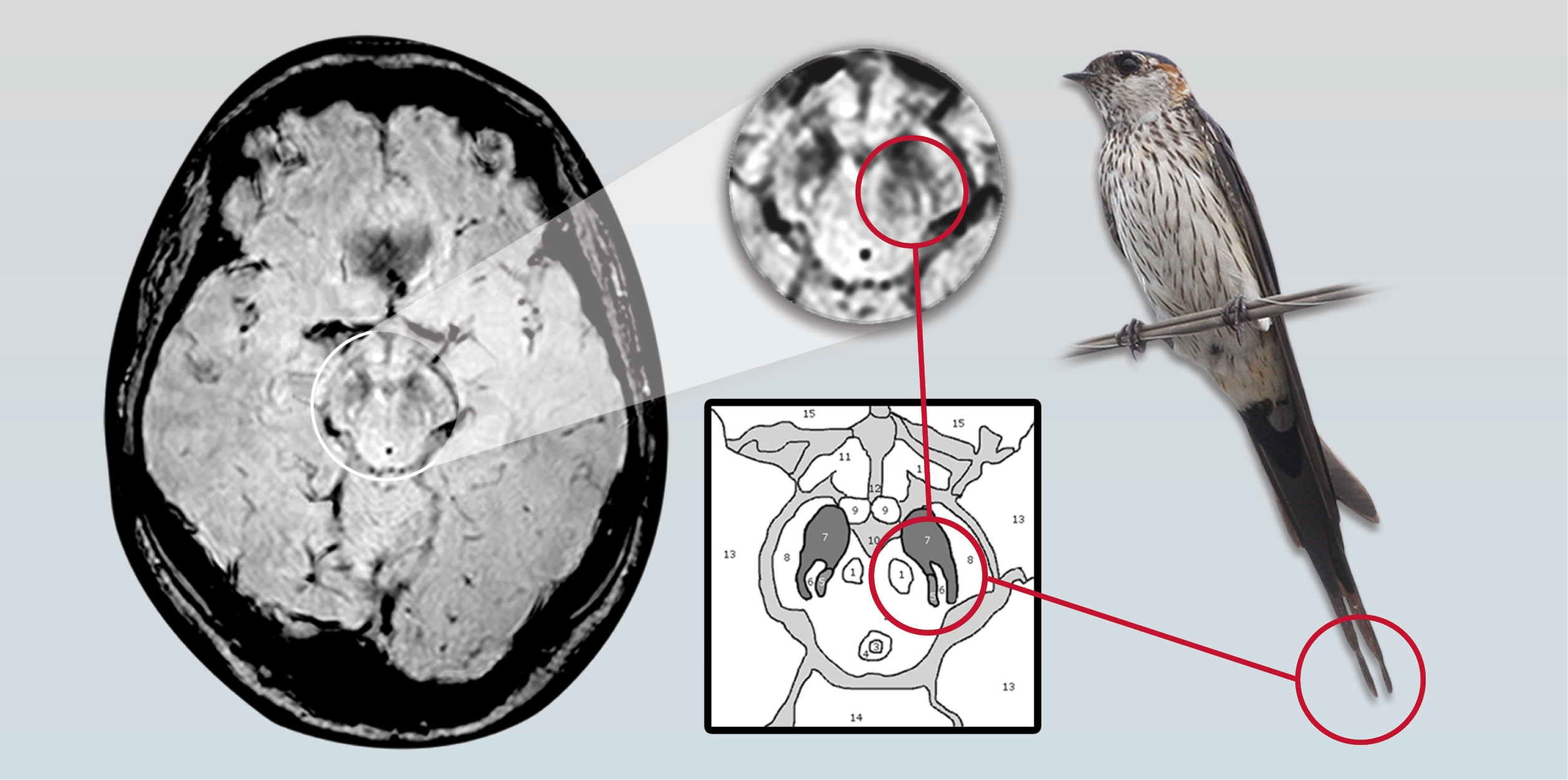
Three MR imaging-based studies have discovered distinctive changes in the substantia nigra (SN), a crescent-shaped mass of cells in the midbrain that normally produces the neurotransmitter dopamine. Parkinson disease patients lose dopamine-producing cells in the SN, leading to problems with motor control among other symptoms. All three studies showed consistent differences in the appearance of the SN in normal patients compared with patients diagnosed with Parkinson disease.
Two of the studies used ultra high-field 7-T MR imaging—cutting-edge technology for research institutions—while the other relied on 3-T MR imaging, which is commonly available in clinical settings.
The initial study was conducted by neuroradiologists, neurologists and physicists of the University of Nottingham in the U.K., and published in the July 2013 edition of Neurology. Led by resesarchers Anna Blazejewska, Ph.D., and Stefan Schwarz, M.D., the team investigated 7-T MR imaging changes to nigrosome-1 of the substantia nigra revealing an oval shaped structure found in healthy patients but absent in patients with Parkinson disease. Researchers were able to correlate their imaging findings to histological findings using immunohisto-chemical staining techniques to demonstrate nigrosome-1.
In a follow-up study by the same research team led by Dr. Schwarz, previous findings were translated to a 3-T MR imaging platform revealing a specific “swallow tail” shape indicating the presence of nigrosome-1—part of the SN that has a dense concentration of dopamine-producing cells. Researchers determined that the split-tail shape was clearly visible in normal patients but not present in the patients with Parkinson disease. The study was published in the April 2014 edition of the journal PLoS ONE.
In the study, researchers reviewed 114 high-resolution images including a prospective case-control study with 10 patients with Parkinson disease and nine control subjects, and a retrospective study of 95 patients (nine with Parkinson disease and 81 without, five non-diagnostic studies excluded). Two raters independently classified subjects into Parkinson disease and non-Parkinson disease groups, according to the absence or presence of nigrosome-1. Researchers accurately classified eight out of nine control subjects and eight out of 10 patients with Parkinson disease in the prospective study, and 77 out of 81 healthy patients and nine out of nine patients with Parkinson disease in the retrospective study.
“Visual radiological assessment yielded a high diagnostic accuracy for Parkinson disease versus an unselected clinical control population,” the authors wrote. “Assessing the substantia nigra … for the typical ‘swallow tail’ appearance has potential to become a new and easy applicable 3-T MRI diagnostic tool for nigral degeneration in Parkinson disease.”
Ultra High-field MR Holds Promise for Parkinson disease
A separate study, published in the June 2014 edition of Radiology, used 7-T MR imaging to distinguish a three-layered organization of the SN in patients without Parkinson disease, as well as distinctive structural changes in the SN of patients with the disease. The research was part of a larger experimental protocol funded by the Italian government to study the clinical impact of ultra high-field MR imaging in diagnosing neurodegenerative diseases.
Researchers at the University of Pisa, led by Mirco Cosottini, M.D., imaged the brains of 38 people, including 17 patients with typical Parkinson disease and 21 healthy people, as well as a brain specimen from a deceased person. (Patients with atypical Parkinson disease or Parkinson disease accompanied by other neurologic or major medical conditions were excluded.) Images revealed a three-layer inner organization of the SN in healthy patients, and loss of that distinctive organization in patients with Parkinson disease.
To determine whether the differences in the 7-T images were a reliable diagnostic marker for Parkinson disease, two neuroradiologists studied images of the healthy SN in eight subjects. Investigators then independently assessed images from the study group, correctly identifying 100 percent of the patients with Parkinson disease and 96 percent of the healthy controls. Each of the raters conducted two blind evaluations one week apart. Results were consistent both with each other and between their own earlier and later assessments.
“We started our work knowing that the inner structure of the SN has never been detected with neuroimaging techniques,” Dr. Cosottini said. “The introduction of the ultra high-field 7-T MR system—the first in our country—gives us a unique opportunity to explore the midbrain in healthy subjects and in patients with Parkinson disease at an extraordinarily high resolution.”
The team is currently studying patients with atypical Parkinson disease symptoms (such as supranuclear palsy, multi-system atrophy and corticobasal degeneration) to evaluate whether the signal changes in the substantia nigra detectable with ultra high-field 7-T MR are unique to Parkinson disease or shared by other conditions, Dr. Cosottini said.
Parkinson Research Continues
Although more research is necessary, the authors believe these findings have the potential to improve early diagnosis of Parkinson disease in the future.
"After confirmation of our findings in further studies, we think the ‘swallow tail’ sign has the potential to change how Parkinson disease is diagnosed and confirmed in the future," Dr. Schwarz said.
Currently, Dr. Schwarz and colleagues are working on the Parkinson disease Magnetic Imaging Repository (PaMIR) project, a multicenter study combining MR imaging and clinical data of early-stage Parkinson disease and healthy controls.
Researchers hope to further advance 3-T MR imaging markers of Parkinson disease, said Dorothee Auer, M.D., Ph.D., the senior author on the PLoS ONE study and head of radiological sciences at the University of Nottingham.
"We will assess the value of MRI-based nigrosome and neuromelanin imaging in tracking Parkinson disease and generate an MR imaging biomarker repository to maximize the clinical benefit of advanced MRI for people with the disease," Dr. Auer said.

Web Extras
- Access the study, “The ‘Swallow Tail’ Appearance of the Healthy Nigrosome—A New Accurate Test of Parkinson Disease,” at Plosone.org.
- Access an abstract of the study, “Visualization of nigrosome 1 and its loss in PD: pathoanatomical correlation and in vivo 7 T MRI,” at Neurology.org.
- Access the study, “MR Imaging of the Substantia Nigra at 7 T Enables Diagnosis of Parkinson Disease,” in the June 2014 issue of Radiology, at RSNA.org/Radiology.