Your Donations in Action: Darrian McAfee
Evaluating a Novel Imaging Tool for Epilepsy


Currently, 30% of patients with epilepsy continue to have seizures despite best medical therapy. One reason is that existing tools have trouble finding the exact area where seizures start.
Lactate (Lac) production is a well-known effect of metabolic reprogramming and is a biomarker in epilepsy. While detection of Lac elevation using conventional magnetic resonance spectroscopy has been extensively studied as a non-invasive method for identifying epileptic brain tissue, the technique suffers from poor spatial resolution, limiting its clinical use in pre[1]surgical mapping for resection.
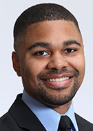
For his 2023 RSNA Research Medical Student Grant project, Darrian McAfee, a medical student at the University of Maryland School of Medicine in Baltimore, and colleagues tested a new imaging method using hyperpolarized 13C magnetic resonance spectroscopic imaging of [1-13C] pyruvate to identify epileptic tissues by detecting increased Lac production.
The researchers found that the method was able to accurately identify elevated Lac production in an in vitro model of chronic hyperactivity and in a gold standard mouse epilepsy model, pentylenetetrazol kindling.
“Our results suggest that this approach has the potential to effectively and non-invasively map epileptic foci and should be further explored as a clinical tool to guide epilepsy resection surgery by identifying epileptic tissue in patients,” McAfee said.
McAfee’s project builds on existing knowledge of the metabolic mechanisms underlying epilepsy and seeks to improve current clinical techniques for more accurate and non-invasive spatial localization of surgical targets.
“Ultimately, it has the potential to enhance the outcomes of resection surgery for epilepsy patients and reduce the burden of presurgical workups,” he said.
Funding from the R&E Foundation helped shape McAfee’s radiology education and aspiration to pursue an academic career. “This project allowed me to explore how radiologic innovations intersect with various medical fields,” he said. “Under the guidance of Drs. Ksendzovsky and Mayer, I gained insight into how radiolabeled molecules, when combined with 3D imaging, can provide precise spatial and temporal information for a wide range of diseases—well beyond the scope of this project.”
For More Information
Learn more about R&E Foundation funding opportunities.
Read our previous Your Donations in Action story.