Deep Learning Helps Speed MRI of the Spine
AI-based method provides excellent image quality with a 70% reduction in exam time


Often ordered to diagnose the causes of pain in the lower back, spine MRI is one of the most common imaging procedures. The ever-increasing number of MRI requests have created an urgent need for examinations that are faster yet still diagnostic.
Deep learning (DL) has shown promise as a tool for cutting examination times for the established MRI technique of turbo spin-echo (TSE) acquisitions. While little is known about the advantages of this DL approach in actual clinical scenarios, the findings of this study are just the latest example of AI-based MRI reconstructions that speed examinations without sacrificing image quality
“The model improves the signal-to-noise ratio and therefore leads to much faster spinal examinations,” said Ahmed E. Othman, MD, professor of neuroradiology at the University Medical Center in Mainz, Germany and senior author of a Radiology study on this topic. “It has already been integrated in a certified clinical sequence protocol and is being used for spinal MR imaging.”
DL Method Interchangeable With Standard TSE
To learn more, researchers in Germany compared DL-reconstructed TSE (TSEDL) with standard TSE in 50 participants. Each participant underwent MRI with each technique. Five experienced radiologists evaluated the images.
DL-reconstructed TSE acquisition was found to be interchangeable with standard TSE for detecting various abnormalities of the spine at MRI. The DL-reconstructed TSE sequences reduced the median total acquisition time from 328 seconds to 100 seconds for a 70% reduction in examination time. No evidence of a difference was found between standard TSE and TSEDL regarding frequency of major findings, overall image quality, or diagnostic confidence.
The findings add to a growing body of research supporting DL-based image reconstruction. In recent years, the researchers for Tuebingen and Mainz University have published similarly promising results from MRI of the hand, wrist, shoulder and head.
“The seamless implementation of the DL-based TSE sequence across a wide range of musculoskeletal MRI examinations underscores its effectiveness and versatility,” said study co-author Judith Herrmann, MD, from Tuebingen University Hospital. “Our ultimate goal is to provide our patients with the most accurate and reliable diagnoses in the shortest possible examination time to improve patient comfort, increase patient throughput and—no less important in the current era—have a positive impact on energy resources.”
Potential future applications include MRI of the upper abdomen, pelvis, prostate, and breast. The researchers are also looking into DL-based MRI reconstructions for whole-body examinations and brain studies.
“The most relevant future accelerated protocol is an ultra-fast brain MRI protocol including five sequences which are being acquired within three minutes,” Dr. Othman said. “Initial analysis reveals very promising results.”
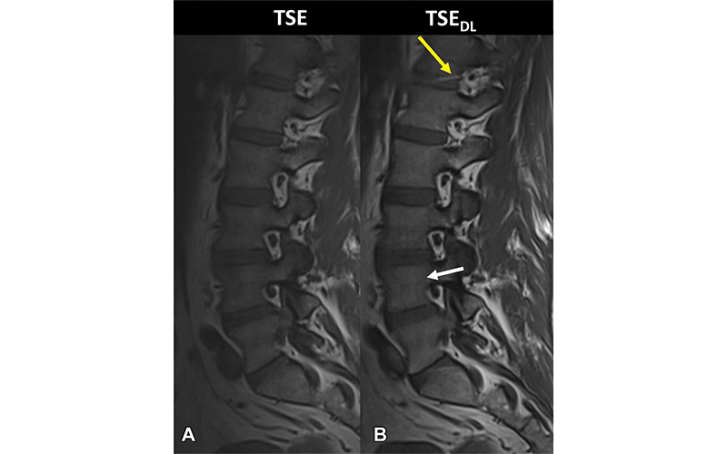
Noncontrast MRI scans at 3 T in a 46-year-old male participant who presented with acute onset of low back pain. (A) Sagittal T1-weighted image acquired with standard fully sampled turbo spin echo (TSE) shows no relevant artifacts. (B) Sagittal T1-weighted image acquired with deep learning–reconstructed TSE (TSEDL) shows characteristic banding artifacts with a streaking pattern aligned with the phase-encoding direction (white arrow). The same image illustrates a white band on the intervertebral disk T12-L1 (yellow arrow), which is an aliasing artifact due to the higher parallel acceleration factor. https://doi.org/10.1148/radiol.212922 © RSNA 2022
Method Has Potential To Improve Imaging Efficiency
While the study results are encouraging, more research is needed to make sure that the DL-based model is generalizable across different patient populations, MRI vendors and imaging sites, according to James T.P.D. Hallinan, MBChB, senior consultant at the Department of Diagnostic Imaging at National University Hospital and Yong Loo Lin School of Medicine, both in Singapore.
In an editorial accompanying the study, Dr. Hallinan wrote that true appreciation of the wider clinical utility of the DL acceleration technique would require validation at other centers with an increased number of participants and range of pathologic findings.
“Clinical studies are necessary to compare standard clinical MRI acquisitions to those obtained with DL to ensure quality and safety are not compromised in the ever-increasing need for speed,” he said.
In addition, Dr. Hallinan said, the implementation of DL algorithms will have to be carried out in close collaboration with regulatory bodies to ensure adherence to existing safety and quality standards.
Dr. Hallinan noted that DL applications are only one potential avenue for reducing MRI acquisition times. Existing MRI acceleration techniques such as compressed sensing and simultaneous multi-slice have a proven track record for reducing MRI acquisition times. These techniques are readily available on most MRI platforms allowing real-time acceleration and are easy to integrate into existing infrastructure, he said. They could also be integrated with DL reconstruction for even faster acceleration.
“What remains clear is that reducing MRI scanning time, while maintaining or improving image quality, will not only reduce costs but have the added benefit of improving patient comfort and satisfaction,” Dr. Hallinan said.
Dr. Hallinan expects these DL-based techniques to continue to improve, driven by advances in model architecture and data augmentation, and the broader availability of training data from multiple sites and vendors to ensure generalizability. He emphasized the importance of making imaging data open source, which will enable international research teams to collaborate and push the boundaries of this field.
For More Information
Access the Radiology study, “Deep Learning Reconstruction for Accelerated Spine MRI: Prospective Analysis of Interchangeability,” and the accompanying editorial, “Deep Learning for Spine MRI: Reducing Time Not Quality."
Read previous RSNA News stories on spinal imaging: