Assessing Aortic Viscous Energy Loss for Valve-related Hemodynamics in Asymptomatic Severe Aortic Stenosis
4D flow MRI-derived energetic markers aid in the clinical management of asymptomatic patients with severe AS

In symptomatic patients with severe aortic stenosis (AS) and those with severe AS and reduced left ventricular ejection fraction (LVEF), treatment with an aortic valve replacement (AVR) is recommended. In up to 40% of patients with severe AS, Doppler echocardiography, which is the standard for imaging and important for surgical decision-making, displays discordant findings among metrics to assess AS severity.
The lack of a strong assessment tool makes it difficult to identify the optimal interventional timing, since early intervention for patients with AS can relieve symptoms, optimize long-term survival and improve quality of life.
Recent advances in four-dimensional (4D) flow MRI indicate that eccentric flow jets and turbulence from a stenotic valve is associated with elevated hemodynamic force toward the ascending aorta and contributes to aortic dilatation. This may be an important metric for interventional planning.
In a Radiology study, Yongshi Wang, MD, from the Shanghai Institute of Cardiovascular Diseases at Zhongshan Hospital, Fudan University, Shanghai, China, and colleagues investigated whether 4D flow MRI could determine AS severity by assessing abnormal blood flow patterns via viscous energy loss (E’L) which is the mechanical energy lost to thermal energy due to frictional forces induced by fluid viscosity. The researchers also wanted to determine if 4D flow MRI could be a valuable complement to conventional Doppler-based metrics for the determination of AS-related prognosis.
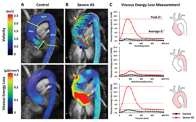
Peak systolic three-dimensional streamlines and viscous dissipation (E′L) in the thoracic aorta of (B) a 53-year-old male patient with severe aortic stenosis (AS) compared with (A) an aortic size-matched healthy control. The direction and propagation of a systolic flow jet in the patient with severe AS was indicated by the dashed black arrow. Regions with high-velocity outflow jets and flow jet impingement exhibited remarkably elevated E′L (yellow arrows), and the presence of pronounced helical flow led to moderate E′L (black arrows). (C) Peak systolic and average E′L increased 10-fold and sevenfold, respectively, in the ascending aorta (AAo) in the patient with severe AS compared with the control and was further elevated along the aortic arch to the descending aorta (DAo). A = anterior, I = inferior, L = left, R = right, S = superior. https://doi.org/10.1148/ryct.220010 ©RSNA 2022
4D Flow MRI Removes Need for Complex Calibrations Using Doppler
In this prospective, single-center study, E’L was measured using 4D flow MRI in the thoracic aortas of 74 consecutive asymptomatic patients with severe AS and preserved LVEF and 23 healthy volunteers. Transvalvular energy loss was assessed based on the energy loss index E’L measured using Doppler echocardiography.
During a median follow-up period of 42 months, 33 patients developed AS-related events including two cardiac-related deaths, six hospitalizations for heart failure, one myocardial infarction that was survived, and 24 aortic valve replacements. Patients with AS-related events exhibited more restricted valve opening, characterized by higher peak transaortic velocity and mean pressure gradient compared to those without an AS-related event.
Researchers noted that complex 3D post-stenotic flow disturbances were accompanied by elevation of irreversible, nonturbulent E’L. Among asymptomatic patients with severe AS and preserved LVEF, peak systolic E’L in the ascending aorta represented inefficient LV energy expenditure and displayed predictive value for clinical outcome. These findings provided proof of concept for 4D flow MRI-based E’L as an appealing parameter to determine AS severity and to improve risk stratification for asymptomatic patients with AS.
“The estimation of the severity of aortic stenosis has been confounded by the discrepancy between Doppler-based metrics and invasive catheterization findings,” Dr. Wang said. “In our study, it was very interesting that peak systolic viscous energy loss in the ascending aorta measured by 4D flow MRI highly correlated with Doppler-based transvalvular energy loss. The introduction of aortic viscous energy loss measurement via 4D flow MRI permits direct estimation and visualization of irreversible, nonturbulent energy cascade dynamics vulnerable to post-stenotic flow disturbances in the aorta. This may eliminate the need for complex calibrations for the Doppler-based assessment of transvalvular energy loss.”
The application of 4D flow MRI is gaining popularity, but technical challenges remain.
“4D flow MRI studies assist cardiac radiologists systematically investigating total irreversible aortic energy loss consisting of both nonturbulent viscous dissipation and turbulent kinetic energy, as well as the interaction between aortic energy loss and LV morphologic and functional changes,” Dr. Wang said. “These findings offer a unique vantage point for cardiac radiologists to consider the application of novel techniques and metrics for advanced clinical evaluation of the pathophysiology of severe AS, especially from the concept of arterial-ventricular coupling.”
For More Information
Access the Radiology: Cardiothoracic Imaging study, “Aortic Viscous Energy Loss for Assessment of Valve-related Hemodynamics in Asymptomatic Severe Aortic Stenosis.”
Read previous RSNA News stories related to cardiac imaging: