International, Multicenter Study Shows Moderate Agreement Among Readers Using LI-RADS
Standardized interpretation and reporting of liver lesions will improve patient care


Multiphase CT and MRI are instrumental in the noninvasive diagnosis and management of liver cancer, which benefits from standardized terminology, technique, interpretation and reporting provided by the Liver Imaging Reporting and Data System (LI-RADS).
“LI-RADS is an important advance in the field of abdominal radiology which has substantially improved standardization and reporting of findings in the liver,” said Sam Galgano, MD, associate professor, vice chair of quality and patient experience at University of Alabama at Birmingham and co-author of a commentary on a recent Radiology study on LI-RADS. “For example, hepatocellular carcinoma is one of the only cancers in the abdomen and pelvis that can be diagnosed noninvasively, but this requires consistency and clear guidelines for what is diagnostic of HCC versus simply suspicious.”
LI-RADS Version 2018 Has Moderate Agreement in Observation and Feature Characterization
The retrospective study assessed reader agreement of LI-RADS in an international, multicenter setting and used scrollable images.
The study included clinically acquired multiphase LI-RADS CT and MRI examinations from six institutions and three countries (United States, South Korea and Colombia) submitted to the coordinating center (University of California San Diego).
Examinations were uploaded to a cloud-based platform and assigned to two of 43 readers. The readers were randomized to ensure they were from separate institutions that were different from the submitting site, eliminating the possibility of familiarity bias. The study included CT and MRI examinations from 484 unique patients being read by radiologists from 33 different institutions, all of whom mostly or almost exclusively read abdominal imaging in their daily clinical practice.
“I have been particularly interested in liver imaging because, unlike most other malignancies in at-risk patients, the diagnosis of hepatocellular carcinoma can be made solely based on imaging. This means that patients can subsequently undergo locoregional therapy or liver transplantation solely based on the radiologist’s read. That is a lot of responsibility, but that also makes these exams particularly interesting and meaningful to interpret,” said author William Hong, MD, assistant professor of radiology at University of California San Francisco.
The study found that LI-RADS generally has moderate agreement for observation categorization and feature characterization. The LI-RADS categories reflect the probability of an observation being an HCC and ranges from LR-1 (definitely benign) to LR-5 (definitely HCC) and feature characterization from imaging can be used to determine the LI-RADS categorization.
“Interreader agreement is key for consistency and for noninvasive diagnosis of HCC. These patients often undergo multiple follow-up exams and consistency between readers is key over time. On a larger scale, for LI-RADS to achieve optimal diagnostic performance, the system needs to work for a wide spectrum of readers at varying degrees of expertise,” Dr. Galgano said. “Overall, the study is an important addition to the field of liver imaging and continues to establish LI-RADS as an essential standardized reporting lexicon and algorithm.”
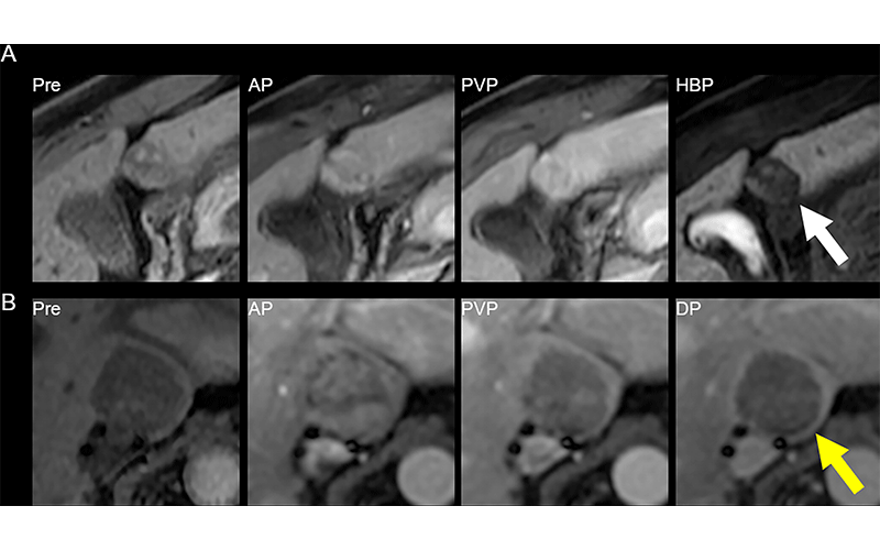
MRI scans show (A) reader disagreement and (B) reader agreement. (A) Gadoxetic acid–enhanced MRI scans in a 56-year-old male patient with cirrhosis secondary to hepatitis C. From left to right: contrast-unenhanced (Pre), arterial phase (AP), portal venous phase (PVP), and hepatobiliary phase (HBP) images. This 21-mm hepatobiliary phase hypointense observation (arrow) was characterized on the clinical read as having nonrim arterial phase hyperenhancement and washout appearance and was categorized as Liver Imaging Reporting and Data System (LI-RADS) category LR-5 (definitely hepatocellular carcinoma [HCC]). The first research reader characterized it as having a targetoid appearance and categorized it as LR-M (probably or definitely malignant, not specific for HCC). The second research reader characterized it as having no major features and paralleling the blood pool and categorized it as LR-2 (probably benign). It was subsequently resected and found to be a well-differentiated HCC. (B) Extracellular contrast–enhanced MRI scans in a 61-year-old female patient with cirrhosis secondary to hepatitis C. From left to right: contrast-unenhanced, arterial phase, portal venous phase, and delayed-phase (DP) images. This 31-mm observation (arrow) in the caudate lobe was characterized on the clinical read as having arterial phase hyperenhancement, washout appearance, and capsule appearance, and was categorized as LI-RADS category LR-5 (definitely HCC). Both research readers also categorized this observation as LR-5. The patient died of intracranial hemorrhage a few months later. https://doi.org/10.1148/radiol.222855 ©RSNA 2023
More Research Needed Using LI-RADS In Clinical Practice
Previous studies assessing the reader agreement of LI-RADS have been limited by factors such as single-center nature, small number of readers, preselected images, and lack of comparison with clinical reads. And although LI-RADS has been extensively validated, that has often been done in academic centers in a research environment. There may be more variability in clinical practice.
“Variability in reporting can lead to confusion for treating physicians; at times, unclear wording can lead to confusion around the patient’s diagnosis, the appropriate level of concern and recommendations for treatment or follow-up,” said Sarah Johnson, MD, assistant professor in the abdominal division of the University of Toronto, Ontario, and author of a second commentary on this Radiology study.
Dr. Hong said he hoped the research shows the immense progress that has been made on LI-RADS since its inception, but also that there is more work to be done.
Dr. Johnson agreed, saying that as more clinically relevant data from real world application of LI-RADS becomes available, the system will continue to improve.
“The LI-RADS system is a work in progress,” Dr. Johnson said. “Standardization of interpretation and reporting of liver lesions will improve patient care. Accurate interpretation by all radiologists reporting liver CT and MRI is a desired outcome. In their study, the researchers have provided further evidence for an ongoing need to improve the standardization of LI-RADS feature assessment and scoring. One can only look forward to the clinical benefit that will result from this achievement.”
For More Information
Access the Radiology study, “A Multicenter Assessment of Interreader Reliability of LI-RADS Version 2018 for MRI and CT.”
Access the Radiology commentary, “LI-RADS Version 2018 for MRI and CT: Interreader Agreement in Real-World Practice.”
Access the Radiology commentary, “Reliability of LI-RADS for MRI and CT: Is Excellence Achievable?”
Read previous RSNA News stories on LI-RADS and liver cancer: