Imaging Advancements Pave the Way to Personalized Medicine in Cervical Cancer
Radiomics, advances in measuring hypoxia show promise in aiding treatment


Not only is cervical cancer one of the most common cancers in women — it’s also one of the deadliest. In fact, cervical cancer ranks as the third deadliest disease among women worldwide.
Chemoradiotherapy remains the standard therapeutic approach for patients with locally advanced cervical cancer, but unfortunately, that therapy doesn’t work for everyone.
According to a new retrospective study published in Radiology: Artificial Intelligence, a better stratification method based on pre-treatment prognosis is needed.
“Such a method could define groups for whom standard therapy is likely to fail and who should be specific cohorts for future clinical research, such as a combination of chemoradiotherapy and target drug or immunotherapy,” said Wei Mu, PhD, co-lead author and a researcher at the H. Lee Moffitt Cancer Center and Research Institute in Tampa, FL.
This method could prevent overtreatment, decrease the physical and economic burden of treatment, and allow patients to start other therapy as soon as possible, Dr. Mu said.
The key to realizing such a method could be radiomics, she said.
“Radiomics extracts a large number of features from radiographic medical images, which have the potential to uncover disease characteristics that can’t be detected by the naked eye,” Dr. Mu explained. “With radiomics, distinctive imaging features are not only related to micro-gene expression patterns, but also to prognosis and therapeutic response, providing valuable information for personalized therapy.”
“Incorporating habitat features into radiomics signatures significantly improved the performance of individualized progression-free and overall survival estimation in cervical cancer patients.”
Wei Mu, PhD
Improving Performance Through Habitat Features
In the Radiology: Artificial Intelligence study, researchers curated PET/CT images and outcomes from 154 patients with locally advanced cervical cancer who underwent chemoradiotherapy from two institutions between March 2008 and June 2016.
The study followed four radiomic steps: tumor segmentation, feature extraction, feature selection and, finally, model construction.
“For tumor segmentation, we first developed a novel level set based on the gradient fields,” Dr. Mu said. “This allowed us to automatically separate the tumor and the nearby bladder and accurately segment the tumor.”
Radiomic features were then extracted from PET and CT images, along with habitat, a physiologically similar area within the tumor, images created by fusing the PET and CT images together — the latter of which is a key differentiator of this study.
“We started by extracting features from the whole tumor,” Dr. Mu explained. “However, we soon found that some patients exhibiting very similar whole tumor-based features had quite different prognoses.”
By dividing the tumor into different habitats and defining a series of features for each habitat, researchers concluded that the features of different habitats play different roles in prognosis.
“A number of PET/CT radiomics studies have proposed signatures for recurrence prediction and prognosis estimation in cervical cancer,” Dr. Mu said. “However, these studies extracted image-derived features independently from PET or CT images, which we have proven to be inferior to fused information.”
By projecting PET data onto the CT images to create a habitat mask, researchers were able to extract the interactive information from images of both modalities. This allowed researchers to better distinguish patients with different prognoses but who showed similar PET or CT features.
“Incorporating habitat features into radiomics signatures significantly improved the performance of individualized progression-free and overall survival estimation in cervical cancer patients,” Dr. Mu explained. Researchers then reduced the dimension of the features according to their stability and predictive ability. Using the LASSO (least absolute shrinkage and selection operator) method to select the most informative features, they constructed the final prognosis prediction models.
According to Dr. Mu, these findings could help characterize different patients quantitatively versus through qualitative descriptions alone and have the potential to help oncologists personalize treatment plans for patients.
“Nomogram models consisting of radiomic signatures and lymph node status obtained from pretreatment PET/CT images and T stage have the potential to identify patients in whom standard chemotherapy is likely to fail and who may need more aggressive treatment in clinical practice,” she concluded.
Researchers now plan to perform further validation using larger prospective cohorts. “We’d also like to export this model to other investigators,” Dr. Mu said.
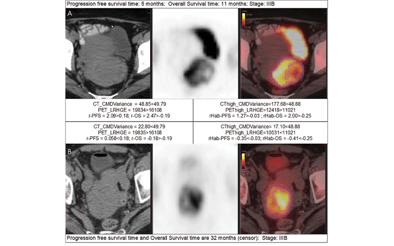
Examples of detailed radiomics signatures. A, B, are the CT, PET, and fusion images of two patients at the same stage but with bad prognosis (progression-free survival [PFS] of 5 months, overall survival [OS] of 11 months, and stage IIIB) and good prognosis (PFS of 32 months, OS of 32 months, and stage IIIB), respectively. The red contours in the fusion images indicate the high metabolic habitat, while the rest of the region in the blue contour is the low metabolic habitat. The cutoffs (49.79, 16108, 48.88, 11021, 0.18, −0.19, −0.03, and −0.25) are obtained using X-tile software on the training dataset. The larger the radiomics signatures (r-PFS, r-OS, rHabPFS, and rHab-OS), the higher the risk of progression or death. CMDvariance = difference variance feature calculated from co-occurrence matrix, LRHGE = long run high gray-level emphasis calculated from run-length matrix, r-OS = radiomic signature for OS obtained with PET plus CT features, r-PFS = radiomic signature for PFS obtained with PET plus CT features, rHabOS = radiomic signature for OS obtained with PET plus CT plus habitat features, rHab-PFS = radiomic signature for PFS obtained with PET plus CT plus habitat features.
Mu et al, Radiology: Artificial Intelligence 2021 ©Radiology: Artificial Intelligence 2021
Measuring Hypoxia in Cervical Cancer
Another area where imaging is playing an increasingly important role is in measuring hypoxia, an adverse factor in cervical cancer.
“Cervical cancer under hypoxic conditions is associated with metastatic progression, extracellular matrix remodeling and poor outcomes,” said Joseph Waller, BA, MS, a medical student at Drexel University College of Medicine in Philadelphia.
Waller co-authored a 2020 review article on the utility in measuring cervical cancer hypoxia via imaging for the British Journal of Radiology.
According to Waller, oxygen measurements of cervix tumors are of paramount importance.
“Not only could these measurements lead to a more accurate prognosis, they are key to developing personalized treatments for patients by taking into account the presence of hypoxic areas that are less responsive to radiation therapy,” Waller said.
Despite this potential, clinically available methods for detecting and evaluating hypoxia during the course of treatment are not yet widespread. To help fill this gap, this review compared methods, summarized strengths and weaknesses, and assessed the best pathways for bringing these methods into clinical practice.
The authors determined that oxygenation level dependent applications of MRI have demonstrated hypoxia-induced radio-resistance and changes in cervix tumor oxygenation from hypoxic therapy.
Furthermore, the hypoxic areas within tumors can be indirectly identified in dynamic contrast-enhanced images, where they generally display low signal enhancement, and in diffusion-weighted images, which demonstrate areas of restricted diffusion that may correlate with hypoxia.
Last, PET used either independently or with other imaging modalities has proven effective in imaging hypoxia through tracers specific for low oxygen levels, such as Cu-ATSM tracers and nitroimidazoles.
“Our findings show that both medical imaging and non-imaging tools like electron paramagnetic resonance oximetry can be clinically used to detect hypoxia in the tumors of patients diagnosed with cervical cancer,” Waller said. “For example, when used to guide radiation and post-treatment surveillance, these methods can result in a much more personalized approach to treatment.”
Moving forward, Waller stressed the need for large clinical trials and further comparative effectiveness research.
For More Information
Access the Radiology: Artificial Intelligence study, “18F -FDG PET/CT Habitat Radiomics Predicts Outcome of Patients with Cervical Cancer Treated with Chemoradiotherapy."
Access the study, “The Clinical Utility of Imaging Methods Used to Measure Hypoxia in Cervical Cancer,” at birpublications.org.