Experts Propose Standardized Criteria for Interpreting, Reporting of CTA-Derived Fractional Flow Reserve Testing
International researchers outline recommendations in a special report in Radiology: Cardiothoracic Imaging

With the increase in use of coronary CT angiography (CTA)-derived fractional flow reserve (FFRCT) to assess patients with stable coronary artery disease (CAD), standardizing the criteria on the interpretation and reporting of results has become critically important.
Consequently, a group of international experts with years of clinical and research experience with FFRCT, has published an expert opinion on a standardized FFRCT interpretation and reporting approach in a special report in Radiology: Cardiothoracic Imaging.
Guidelines now recommend CTA as a valuable first line test in patients suspected of having stable CAD. However, according to authors of the report, led by Bjarne L. Norgaard, MD, PhD, Department of Cardiology, Aarhus University Hospital, Aarhus, Denmark, CTA could overestimate stenosis severity, particularly in the event of coronary calcification.
Moreover, CTA is a strictly anatomic test, making it a poor discriminator of lesion-specific ischemia as determined by invasive fractional flow reserve (FFR), which is the reference standard for decision-making on coronary revascularization.
“Thus, when compared to conventional non-invasive functional testing such as stress echo, SPECT and PET for first-line assessment of stable chest pain patients, CTA may be associated with increased downstream utilization of invasive catheterization and revascularization,” Dr. Norgaard said.
He pointed out that guidelines recommend CTA be used in patients with a lower likelihood of CAD, and that additional traditional non-invasive or invasive functional evaluation should be performed in the event of a positive CTA result.
FFRCT is a new functional test based on standard acquired CT datasets, without the need for additional radiation or medication, providing information on computational FFR in all aspects of the coronary tree. “An abundance of data has demonstrated that FFRCT has high diagnostic accuracy when compared to FFR, and in several reports FFRCT, showed promise as a valuable test in clinical practice,” Dr. Norgaard said. “For example, several studies indicate that FFRCT, may offset the higher downstream utilization of invasive catheterization by CTA and favorably influence clinical outcomes.”
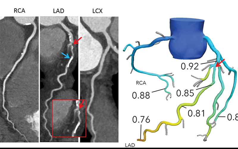
Left: Interpretation of FFRCT results in a 65-year-old woman with typical angina. Agatston score, 333. Left: Coronary CT angiography curved multiplanar reconstructions demonstrate a 50%–69% proximal left anterior descending artery (LAD) stenosis (red arrow) and in the mid-LAD, nonobstructive diffuse disease. The blue arrow indicates where the lesion-specific FFRCT value was assessed. Right: In the FFRCT three-dimensional model, the FFRCT value 16 mm distal to the stenosis was 0.85, indicating that the lesion did not cause significant pressure loss. However, FFRCT was significantly low (0.76) in the terminal vessel segments.
Norgaard, et al, Radiology: Cardiothoracic Imaging 2020 @RSNA 2020
Standardizing Criteria Approach for Interpretation, Reporting
While there has been much focus on the diagnostic performance and potential clinical utility of FFRCT in patients with moderate CAD, Dr. Norgaard noted that there has been less focus on the interpretation, reporting and integration of FFRCT results into routine clinical practice.
“A broadly adopted standardized FFRCT interpretation and reporting approach providing rich and consistent information may facilitate more appropriate clinical implementation and stimulate further high-quality research,” writes Dr. Norgaard and his colleagues in their report. Therefore, their objective was to propose standardized criteria for FFRCT interpretation and reporting that could be applied in clinical practice and for clinical research.
Dr. Norgaard and colleagues issued several recommendations.
• Use the FFRCT value 10-20 mm distal to the stenosis for decision-making.
• FFRCT values that are remote from a specific coronary lesion may be clinically relevant but should not be used alone to assess the use of invasive management of that lesion.
• A dichotomous interpretation strategy for clinical decision-making should be considered only in lesions with FFRCT >0.80 or ≤0.75, whereas additional risk stratification (especially regarding symptoms) information is needed in patients with FFRCT ranging between 0.76 and 0.80.
Moreover, the results of FFRCT must always be evaluated in its clinical context, taking into account patient symptoms, the coronary anatomy, and suitability of revascularization.
The authors stressed that FFRCT analyses should not be performed in all patients. For example, the utility and diagnostic performance of FFRCT has only been studied in patients suspected of having stable CAD, they said. “The use of FFR in patients with stents or bypass grafts, microvascular dysfunction, prior myocardial infarction or suspected or known acute coronary syndromes cannot be recommended at this point,” the authors write. “More studies are needed.”
Currently, large-scale multicenter randomized studies are underway to define the exact role CTA will play with selective FFRCT testing in clinical practice, compared to usual care — including the use of conventional functional testing modalities such as stress echo, SPECT and PET.
“From these outcome studies we will learn more regarding the optimal FFRCT interpretation strategy,” Dr. Norgaard said.
For More Information
Access the Radiology: Cardiothoracic Imaging Special Report, “Coronary CT Angiography-derived Fractional Flow Reserve Testing in Patients with Stable Coronary Artery Disease: Recommendations on Interpretation and Reporting.”