Quantitative Mapping of Salivary Gland Tumors Promising in Determining Malignancy
Researchers Analyze and Classify Pathology-Proven Salivary Gland Tumors Using T1 and T2 Heat Mapping.

A University of Chicago (UC) researcher has developed a promising, non-invasive technique designed to evaluate the potential malignancy of preoperative salivary gland tumors using quantitative T1 and T2 mapping.
“Although preliminary, the findings raise the exciting possibility of using T1 and T2 mapping as a diagnostic aid, capable of differentiating between benign and malignant tumors, which at this point can only be accomplished with tissue biopsy,” said study author Annie Xiao, BS, a medical student at UC who plans to pursue cancer research as a physician-scientist.
Under the supervision of Daniel Thomas Ginat, MD, director of head and neck imaging at UC, Xiao completed the research with the aid of a 2018 RSNA Medical Student Research Grant.
“We wanted to investigate if there were any features identifiable on MRI that would suggest whether the tumors were benign or malignant,” Xiao said.
Salivary gland lesions comprise approximately six percent of head and neck neoplasms and present a diagnostic challenge for physicians. Current diagnostic modalities such as high-resolution ultrasound (US) and fine needle aspiration cytology (FNAC) either do not provide reliable accuracy or present safety risks for patients without offering objective tumor assessment, Xiao said.
“High-resolution US features of some benign and malignant masses have been shown to overlap, rendering the two indistinguishable,” she said. “And invasiveness, controversial diagnostic value, and concerns about the risk of tumor cell seeding have prevented FNAC from achieving widespread clinical acceptance.”
Researchers sought to fill that unmet need for an effective, non-invasive tool to differentiate benign and malignant parotid gland neoplasms, Dr. Ginat said. “Our goal was to make the interpretation of parotid tumors more objective, as imaging enters the age of AI and other forms of quantitative, computer-assisted interpretation,” Dr. Ginat said.
Read the study process and results in the May print issue of RSNA News.
Grants in Action
NAME:
Annie Xiao, BS
GRANT RECEIVED:
RSNA Research Medical Student Grant (2018)
STUDY:
“Quantitative T1 and T2 Mapping of Salivary Gland Tumors: MRI Correlation with Histopathological Features of Cellularity and Malignancy.”
CAREER IMPACT:
“This grant program has been beneficial to my development as a medical student clinical researcher. Through this experience, I have strengthened my problem-solving and critical thinking skills and become more knowledgeable about concepts in radiology pertinent to my project,” Xiao said.
CLINICAL IMPLICATION:
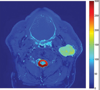
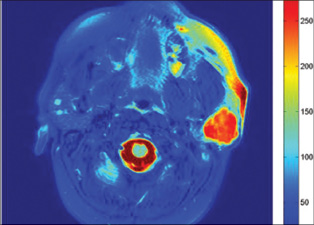
“With a modest sample size, we are able to show drastic differences in T2 values between pleomorphic adenomas as a group and all other salivary gland tumors. The quantitative T2 heat maps developed in this study present a powerful visual tool to aid radiologists in their interpretation of patient MR scans.”