Alternatives to Gadolinium-Based Contrast Agents Show Potential
Radiology researchers study xenon gas and iron-based contrast agents


Gadolinium-based contrast agents have been used for diagnosis and treatment guidance on more than 100 million patients worldwide over the past 25 years.
And for good reason, said Eyk Schellenberger, MD, professor of imaging and head of the Molecular Imaging Group, Institute of Radiology, Charitė — Univerisitätsmedizin Berlin, Germany. Gadolinium-based contrast agents possess a unique electronic structure that make them strongly paramagnetic, and therefore extraordinarily useful as an MR contrast agent.
But over the years concerns have arisen about the safety of gadolinium-based contrast agents. And recently, several preliminary studies showed the presence of residual gadolinium concentrations in brains of patients who had no history of kidney disease.
In July 2017, the European Medicines Agency (EMA) confirmed its previous conclusion that there is convincing evidence of gadolinium deposition in brain tissues following the use of gadolinium contrast agents. The agency recommended restrictions on the use of linear gadolinium agents.
In September 2017, the FDA Medical Imaging Drugs Advisory Committee (MIDAC) recommended adding a warning to labels about gadolinium retention for gadolinium-based contrast agents used during MRI.
Shortly after the FDA’s recommendation, RSNA issued a statement explaining that patients should not be unnecessarily deprived of crucial, sometimes life-saving medical data from gadolinium contrast-enhanced MRI.
“At the same time, the potential risk associated with residual gadolinium concentrations in the brain should be taken into consideration,” RSNA advised in a position statement. “This risk must be weighed against the clinical benefit of the diagnostic information or treatment results that MRI or [magnetic resonance angiography] may provide for each patient.”
“Whether these accumulations are dangerous or not is not clear yet,” Dr. Schellenberger said. “Consequently more research is necessary and potentially safer alternatives are desirable.”
Iron-Based Contrast Shows Potential
Researchers such as Dr. Schellenberger are investigating the possibility of alternatives to gadolinium-based contrast agents.
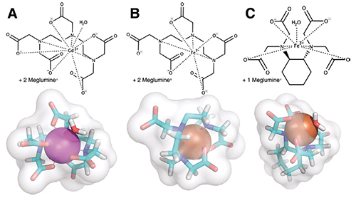
In a recent Radiology study published online, Dr. Schellenberger and colleagues studied iron-based contrast agents as a possible alternative. While gadolinium is a trace element that does not occur in the body in relevant amounts, ferric iron (Fe3+) — which also has a relatively high paramagnetism — is a substance known — and needed — by the body, Dr. Schellenberger explained.
“The body has a dedicated system for safe uptake, transport and storage,” he said. “Moreover, the stabilities of chelates of iron are generally several orders of magnitude higher than those of gadolinium. Thus, the amounts of iron potentially released from low molecular weight iron-based contrast agents (IBCA) should be similar or even lower than those released from gadolinium-based contrast agents.”
The researchers synthesized low-molecular-weight iron chelates of trans-cyclohexane diamine tetraacetic acid and pentetic acid and compared their T1 contrast effects to the commercial gadolinium-based contrast agent, Magnevist, in a breast cancer mouse model.
“The study showed that two different iron chelates, when dosed slightly higher — two- and five-fold — deliver the same results in typical applications such as DCE-MRI or MR angiography as Magnevist can,” Dr. Schellenberger said.
Xenon Gas Shows Promise
Another recent development for imaging tissue perfusion with MRI without the use of gadolinium, is inhaled xenon gas, which was the focus of another study recently published in Radiology online.
In the study, the team of Jim M. Wild, PhD, professor of MR physics, POLARIS group, Academic Radiology, University of Sheffield, Sheffield, U.K., performed in vivo imaging with inhaled hyperpolarized xenon 129 (129Xe) MRI, an injection-free means of imaging the perfusion of cerebral tissue in healthy participants.
“Xenon is a noble gas that can be safely inhaled,” Dr. Wild said. “And we can boost the MRI signal from 129Xe using a laser hyperpolarization process, which means that with small quantities of inhaled gas we get a large MR signal.”
The Sheffield team also demonstrated that they could image the uptake of inhaled xenon gas from the air spaces in the lungs, in to the bloodstream and then into the brain tissue itself across the intact blood-brain barrier.
“Existing MRI contrast agents don’t do this — they stay in the intravascular space,” Dr. Wild said. “So, we have a new way of looking at brain tissue perfusion and blood-brain barrier gas exchange with inhaled xenon brain MRI.”
This method has obvious clinical implications, since it involves a means of imaging brain perfusion without having to inject any contrast agent. The contrast comes from an inert gas inhaled in a modest dose of approximately a liter, which is cleared from the body by breathing out.
“More interesting, from a scientific perspective and to understand brain disease, is the ability to look at blood-brain permeability and tissue-blood physiology in the brain,” he added. “Short of PET tracers like oxygen-15 and xenon-ehnahced brain CT, I don’t think there is an MRI contrast agent out there that can do that.”
As for image quality, Dr. Wild and colleagues were able to capture images that were on a par with PET scans, but not of a gadolinium-enhanced brain perfusion MR image.
“That is not to say we can’t get there,” he said.

Web Extras
- Access the studies, “Low-Molecular-Weight Iron Chelates May Be an Alternative to Gadolinium-based Contrast Agents for T1-weighted Contrast-enhanced MR Imaging,” and “Imaging Human Brain Perfusion with Inhaled Hyperpolarized 129Xe MR Imaging1,” at pubs.rsna.org.