Researchers Establish Target Doses for Pediatric Chest CT
Radiology researchers have established diagnostic reference ranges (DRRs) for pediatric chest CT, potentially enabling physicians and technologists to better manage pediatric doses while maintaining image quality.
A relatively new quality improvement tool, DRR, provides a minimum estimated patient radiation dose, below which image quality may not be diagnostic, and an upper estimated patient dose, above which the dose may be larger than necessary.
In new Radiology research, lead author Keith J. Strauss, MSc, and colleagues determined DRRs for pediatric chest CT based on the size of a patient’s chest. In addition, they developed Pediatric Dose Reduction Factors (PDRFs), which allow an estimate of the necessary radiation dose for an individual patient’s chest for each unique CT scanner of a facility. The online tool is scheduled to be available on the Image Gently website (ImageGently.com) by April 1 2017.
While diagnostic reference levels (DRLs) are used as benchmarks for radiation protection, DRRs create a range by establishing a maximum (the same as the DRL) and a minimum recommended estimated patient dose.
“This concept addresses two concerns: appropriate dose level and quality of the image. The PDRF allows a department to set up target dose values for chest CT exams on any size patient on an as-needed basis,” said Mr. Strauss, Associate Professor at the University of Cincinnati School of Medicine.
Co-author Alexander J. Towbin, MD, the Neil D. Johnson Chair of Radiology Informatics at the University of Cincinnati School of Medicine, was instrumental in developing the data allowing the lower bound of the DRR range, Mr. Strauss said.
In the multi-center study, Mr. Strauss and colleagues analyzed three CT dose indexes — CT dose index (CTDI), dose length product (DLP) and a relative newcomer, size specific dose estimate (SSDE) — of 581 patients younger than 21 years old who underwent CT examinations between July 2012 and June 2013. Data was stored in the American College of Radiology (ACR) Dose Index Registry. Five hospitals formed a consortium, Quality Improvement Registry in CT Scans in Children (QuIRCC)to conduct the study.
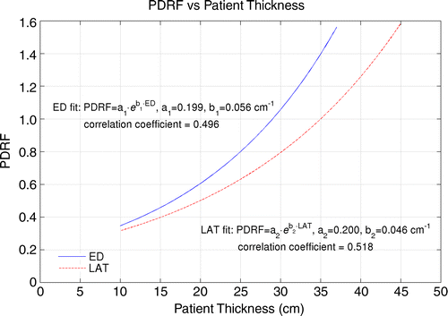
Above: (a–b) Scatterplots of SSDEs with fitted exponential curves as a function of patient size show (a) effective diameter (ED) and (b) lateral dimension (LAT). Coefficients of linear fits allow calculation of average SSDE as a function of patient effective diameter or lateral dimension. (c) Graph shows two normalized fitted curves of SSDE in a and b to SSDE of average-sized adult chest: 35 cm (14) and 29 cm for lateral dimension and effective diameter, respectively (11). Coefficients for two normalized fits based on effective diameter and lateral dimension of patient allow establishment of PDRFs if adult SSDE for unenhanced chest CT examinations for each scanner and size of individual pediatric patient is known. ©RSNA 2017. All rights reserved. Reprinted with permission.
The authors developed DRRs after analyzing the image quality of a subset of 111 CT examinations to validate image quality at the lower range. The PDRF is simply the SSDE for a pediatric patient divided by the SSDE of adult patients at the QuIRCC hospitals.
Pediatric Dose Reduction Factor Works on Any CT Scanner
The proposed PDRFs provide an acceptable range of SSDEs that should help radiology technologists better manage both radiation dose and image quality, Mr. Strauss said.

The table above provides the results of the image quality analysis. The yes (diagnostic) and no (nondiagnostic) ranking determined by each reviewer (A–F) for each set of lung or soft-tissue reconstructions for each of the 111 subjects is provided. Three patients (3%) were scored as nondiagnostic for both soft-tissue and lung reconstructions. Two subjects (2%) and three subjects (3%), respectively, had lung and soft-tissue reconstructions with images of insufficient quality for diagnosis. © RSNA 2017. All rights reserved. Reprinted with permission.
“If I am a technologist unsure of the technique to use on a child, I can measure the lateral dimension of the patient and check a PDRF chart to determine the reduction factor to obtain the pediatric dose compared to the adult dose used at the hospital on a given scanner,” Mr. Strauss said.
The PDRFs should be especially helpful in adult hospitals, which conduct the majority of CT scans on pediatric patients, Mr. Strauss said. Radiology technologists who might not be familiar with pediatric protocols can access the PDRF chart online or post the chart on the scanner’s control console and adjust the patient’s radiation dose accordingly, he said.
Even if a hospital has multiple different CT scanners, Mr. Strauss stresses that the same PDRF chart can be used on any of them, since the adult dose — which is determined for each machine based on its unique design and characteristics— is the starting point for calculating the pediatric dose.
“That’s the beauty of this technique,” Mr. Strauss said. “The PDRF factor should not change over time. Over time as technical improvements to scanners and techniques are developed, these improvements will be incorporated into the facility’s unique adult doses — the starting point for all estimates of appropriate pediatric radiation doses.”
In 2013 Radiology research, the QuIRCC developed PDRFs for abdominal CT that are available on the Image Gently website.
Web Extras
- Access the 2017 Radiology study, “Pediatric Chest CT Diagnostic Reference Ranges: Development and Application,” at pubs.rsna.org
- Access the 2013 Radiology study, “Diagnostic Reference Ranges for Pediatric Abdominal CT,” at pubs.rsna.org
- Access Image Gently at imagegently.org