Methylene Blue Shows Promise for Improving Short-Term Memory


Duong
Duong

Low-dose methylene blue can increase functional MRI activity during sustained attention and short-term memory tasks and enhance memory retrieval, according to new Radiology research.

Pavel Rodriguez, MD, of the Research Imaging Institute in San Antonio, Texas, and colleagues studied 26 subjects to investigate the sustained-attention and memory-enhancing neural correlates of the oral administration of methylene blue in the healthy human brain.

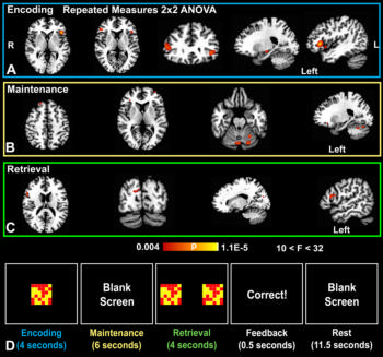
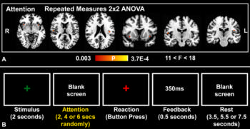
The administration of methylene blue increased response in the bilateral insular cortex during a psychomotor vigilance task and functional MRI response during a short-term memory task involving the prefrontal, parietal and occipital cortex, according to results. Methylene blue was also associated with a 7 percent increase in correct responses during memory retrieval.

“The results support the notion that methylene blue enhances memory performance and functional MRI activity in brain regions associated with a visuospatial short-term memory task. This work provides a neuroimaging foundation to pursue clinical trials of methylene blue in patients undergoing healthy aging and those with cognitive impairment, dementia, or other conditions who may benefit from drug-induced memory enhancement,” the authors write.
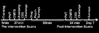
Schematic shows the timeline of functional MR imaging and surveys
Figure 1. Schematic shows the timeline of functional MR imaging and surveys, including psychomotor vigilance task (PVT) and delayed match-to-sample(DMTS) task.
Psychomotor vigilance Task
Figure 2. Psychomotor vigilance Task. A, Repeated-measures analysis of variance (ANOVA) results of the attention phase of the psychomotor vigilance task superim¬posed to a standard brain template (colors indicate P values of significant voxels, cluster-based P ˂ .05; cluster size, K ≥ 10) shows positive drug X time interactions in favor of a methylene blue effect in the bilateral anterior and posterior insular cortices (n = 26, with 24 degrees of freedom). B, Representative block from the psychomo¬tor vigilance task shows the stimulus, attention, reaction, feedback, and rest phases as demarcated by set times.
Delayed match-to-sample task. Repeated-measures ANOVA results of the delayed match-to-sample task
Figure 3. Delayed match-to-sample task. Repeated-measures ANOVA results of the delayed match-to-sample task (colors indicate P values of significant voxels, cluster-based P ˂ .05; K ≥ 10) superimposed to a standard brain template. A, Probability map overlays show positive drug X time interactions in favor of a methy¬lene blue effect in the bilateral inferior frontal gyri during the encoding phase, B, in the right superior frontal gyrus, left middle frontal gyrus, and posterior cerebellum during the maintenance phase, and, C, in the right inferior frontal gyrus and cuneus during the retrieval phase (n = 26, with 24 degrees of freedom). D, Representa¬tive blocks from the delayed match-to-sample task show the encoding, maintenance, retrieval, feedback, and rest phases as demarcated by set times.

Web Extras