Insomnia Linked to Damage in Brain Communication Network


Li
Li

White matter (WM) tracts related to regulation of sleep and wakefulness, and limbic cognitive and sensorimotor regions, are disrupted in the right brain in patients with primary insomnia. The reduced integrity of these WM tracts may be because of loss of myelination, according to new research published in the journal, Radiology.

Shumei Li, M.S., of Guangdong No. 2 Provincial People’s Hospital in Guangzhou, China, and colleagues compared changes in diffusion parameters of WM tracts from 23 primary insomnia patients and 30 healthy control (HC) participants, and the accuracy of these changes in distinguishing insomnia patients from HC participants was evaluated.

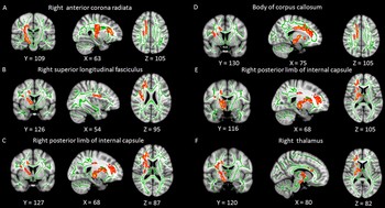
Primary insomnia patients had lower fractional anisotropy (FA) values mainly in the right anterior limb of the internal capsule, right posterior limb of the internal capsule, right anterior corona radiata, right superior corona radiata, right superior longitudinal fasciculus, body of the corpus callosum, and right thalamus.

“Our study suggests that primary insomnia is characterized by altered structural connectivity related to regulation of sleep and wakefulness, particularly involving limbic cognitive function and sensorimotor regions,” the authors write.
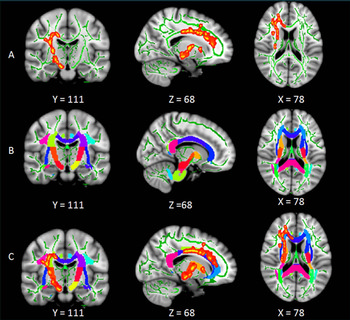
Tract-based analysis for whole WM tracts. A, Significant differences in WM detected from analysis of tract-based spatial statistics.
Figure 1. Tract-based analysis for whole WM tracts. A, Significant differences in WM detected from analysis of tract-based spatial statistics. Results are shown overlaid on the Montreal Neurologic Institute 152-T1 template and the mean FA skeleton (green). B, The Johns Hopkins University International Consortium of Brian Mapping Diffusion Tensor Imaging 81 atlas that covers the entire skeletons of major WM tracts is shown overlaid on the Montreal Neurologic Institute 152-T1 template. C, The overlaid region of significant clusters obtained from tract-based spatial statistics analysis and the Johns Hopkins University International Consortium of Brian Mapping Diffusion Tensor Imaging 81 atlas. The overlapping WM labeling from the Johns Hopkins University International Consortium of Brian Mapping Diffusion Tensor Imaging 81 atlas was used to extract the whole WM and to calculate the mean FA value that represents the integrity of the whole tract.
Significant WM clusters obtained from tract-based spatial statistics between primary insomnia and HC groups.
Figure 2. Significant WM clusters obtained from tract-based spatial statistics between primary insomnia and HC groups. Green represents the mean FA skeleton and red-yellow shows clusters with a significant FA reduction in the primary insomnia group (P ˂ .05, family-wise error correction). Underlying gray-scale images are the Montreal Neurologic Institute 152-T1 template. Each panel is the best axial, coronal, and sagittal view that shows the specific WM tract.
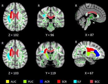
Distribution of the six whole WM tracts in the brain. ALIC = anterior limb of the internal capsule, ACR= anterior corona radiata, BCC = body of the corpus callosum, PLIC = posterior limb of the internal capsule,R = right side of the brain, SCR = superior corona radiata, SLF = superior longitudinal fasciculus.
Figure 3. Distribution of the six whole WM tracts in the brain. ALIC = anterior limb of the internal capsule, ACR= anterior corona radiata, BCC = body of the corpus callosum, PLIC = posterior limb of the internal capsule,R = right side of the brain, SCR = superior corona radiata, SLF = superior longitudinal fasciculus.

Web Extras