CT Allows Nonsurgical Management of Some Lung Nodules
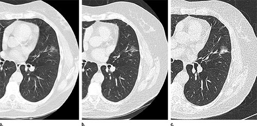
People who have nonsolid lung nodules can be safely monitored with annual low-dose CT screening, according to a new study published online June 23 in Radiology. Researchers say the findings could help spare patients from unnecessary surgery and additional imaging.
Nonsolid nodules are commonly visible on CT scans of the chest, but management of them is challenging, researchers say.
"Nonsolid nodules could be due to inflammation, infection or fibrosis, but could also be cancerous or a precursor of cancer," said study co-author Claudia I. Henschke, Ph.D., M.D., from the Department of Radiology at Icahn School of Medicine at Mount Sinai in New York City. "For screening, we have to define which nodules need further workup and how quickly we have to do that workup."
In the new study, Dr. Henschke and colleagues analyzed results from 57,496 participants in the International Early Lung Cancer Program (I-ELCAP), a major worldwide initiative focused on reducing deaths from lung cancer. Patients underwent baseline and annual repeat screenings, and researchers evaluated the prevalence of nonsolid nodules and their effect on long-term outcomes.
A nonsolid nodule was identified in 2,392 (4.2 percent) of the baseline screenings, and further analysis led to the diagnosis of 73 cases of cancer. A new nonsolid nodule was identified in 485 of 64,677 annual repeat screenings, or 0.7 percent, and 11 were diagnosed with Stage I cancer. Surgery was 100 percent curative in all cases, with a median follow-up since diagnosis of more than six years.
The nonsolid nodule developed a solid component—a warning sign of invasive cancer—in 22 cases prior to treatment. However, the median transition time from nonsolid to part-solid was more than two years. No cancers occurred in new nodules 15 millimeters or larger in diameter.
Findings suggest that nonsolid nodules of any size can be safely followed with low-dose CT at 12-month intervals to assess a potential transition to part-solid.
"The results show that if we see a nonsolid lung nodule of any size, we can tell people to come back in one year for another CT," Dr. Henschke said. "These findings are important for reducing unnecessary CT scans and possible biopsies or surgery in programs of CT screening for lung cancer."

Web Extras
- Access the study, “CT Screening for Lung Cancer: Nonsolid Nodules in Baseline and Annual Repeat Rounds,” at pubs.rsna.org/doi/full/10.1148/radiol.2015142554