Survivors of Childhood Cancer: Risk of Future Disease, Screening Needs and the Role of Radiology
Following successful treatment of primary cancer, patients may gain a false of security and miss future signs of disease

As treatment for childhood cancers continues to improve, so too has the chance for long-term survival. But as childhood cancer survivors enter adulthood, they face an increased risk for developing new conditions related to their prior cancer therapy.
“Because developing tissue in young children is particularly radiosensitive and prone to DNA damage or modification, those treated with radiotherapy at a young age are at an increased risk of developing cancers later in life,” said Yiming Gao, MD, associate professor and program director, breast imaging fellowship at NYU Grossman School of Medicine. “While the actual risk will vary by age of exposure, body part exposed, type of radiation, concurrent chemotherapy and individual patient genetic make-up, the heightened risk persists well into adulthood.”
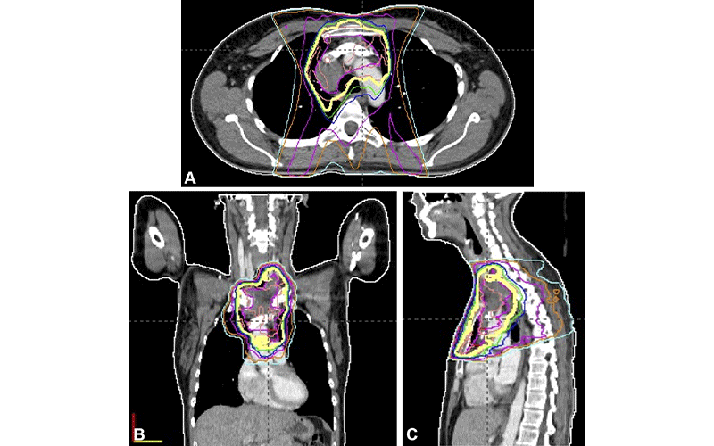
Radiation-Induced Breast Cancer Risk
According to a recent RadioGraphics paper authored by Dr. Gao and colleagues, women who survived childhood cancers are at high risk for developing breast cancer later in life. In fact, by the age of 50, one in three women treated with chest radiation therapy at a young age will be diagnosed with breast cancer, and the cumulative breast cancer incidence in survivors is 13%–20% by age 40–45 years. Mortality rates after breast cancer among survivors of cancer in childhood are higher than those in women with de novo breast cancer. There is also a higher incidence of nonsynchronous second breast cancer among these survivors.
“Advances in radiation therapy continue to curtail exposure, but the risk of a second cancer has no known dose threshold and a long latency period,” Dr. Gao explained.
An Increased Risk for Meningioma
Radiation therapy during childhood can also increase the risk for developing a number of conditions impacting the central nervous system as an adult. For example, according to a 2021 RadioGraphics article, adult survivors of childhood cancer who were exposed to cranial radiation are at an increased risk for meningioma.
“Although the absolute risk of radiation-induced meningiomas is not known, the latency period can be more than 20 years and the incidence continues to increase even after several decades and does not plateau over time,” said Masaki Katsura, MD, PhD, a radiologist at the University of Tokyo.
The article, which Dr. Katsura co-authored, cites a cohort study of over 4,000 childhood cancer survivors exposed to cranial radiation where the cumulative risk of subsequent meningioma was 5.6% by the time the patient was 40 years old.
Furthermore, cohort studies from the Childhood Cancer Survivor Study and British Childhood Cancer Survivor Study demonstrate that the dose of radiation exposure to the cranium has a linear relationship with the risk of developing radiation-induced meningiomas.
“Radiologists are well positioned to contribute to the wider education of patients and referring physicians, which will be paramount in ensuring consistent long-term prevention in all survivors.”
YIMING GAO, MD
The Importance of Screenings
While advanced techniques to limit the field of exposure and minimize radiation dose have helped lower future risk of secondary radiation induced cancer, it is impossible to eliminate long-term cancer risk.
“Although the planning and delivery techniques of radiation therapy have evolved substantially during the past few decades, the structures surrounding the target lesion are inevitably exposed to radiation,” Dr. Katsura explained.
This underscores the importance of screenings.
“Breast cancer screening is cost effective in the general population due to the underlying prevalence of disease and proven mortality benefit of screening,” Dr. Gao said. “In survivors of childhood cancer, this calculus of cost-benefit is even more favorable, as the likelihood of cancer is higher and the average age of developing a second cancer in this group is younger, meaning more life-years saved.”
Unfortunately, breast cancer screening attendance among childhood cancer survivors is poor. Nearly one-half of survivors younger than 40 who previously underwent chest radiation therapy never undergo mammography. Among women ages 40–50 years, only 52% undergo regular mammographic screening, while adherence to MRI screening is even worse.
“The long latency of secondary cancer development after successful treatment of primary childhood cancer may create a false sense of security,” Dr. Gao added. “Continuity of care may also be disrupted when transitioning from pediatric to adult medical care, with timely screening often overlooked.”
Ensuring Consistent Long-Term Prevention in All Survivors
With this in mind, radiologists should promote pertinent long-term screening recommendations when engaged in care for childhood cancers or when encountering adult survivors later in clinical care, either as part of the multi-disciplinary team or when directly communicating with patients and their care providers.
“Radiologists are well positioned to contribute to the wider education of patients and referring physicians, which will help promote and ensure more consistent long-term prevention in all survivors,” Dr. Gao said.
Radiologists should also maintain familiarity with the patient’s timeline and expected imaging appearances after radiation therapy. Knowledge about the radiation treatment plan, number of normal structures included, location of the target lesion and amount of time elapsed since radiation therapy are all particularly important at follow-up imaging.
“As treatment options in oncology continue to expand and long-term survival increases, diagnostic radiologists should be familiar with the patient’s timeline and expected imaging appearances after radiation therapy to avoid pitfalls in image interpretation,” Dr. Katsura concluded.
For More Information
Access the RadioGraphics article, “Breast Cancer Screening in Survivors of Childhood Cancer."
Access the RadioGraphics article, “Recognizing Radiation-induced Changes in the Central Nervous System: Where to Look and What to Look For."
Read previous RSNA News articles on imaging and cancer: