Photon-Counting CT Can Evaluate Lung Function
Substantial improvement seen in contrast-to-noise ratio, spatial resolution of pulmonary blood volume images

New CT technology allows for a comprehensive, simultaneous evaluation of lung structure and function, something not possible with standard CT, according to a study published in Radiology.
Chest CT is the imaging method of choice for analyzing lung disease and tracking changes over time. However, CT studies of lung function and perfusion require dedicated protocols that cannot be combined.
Researchers in Germany and the Netherlands developed a chest imaging protocol that yields information on structure and function of the lungs as an all-in-one procedure. The protocol uses photon-counting CT technology that enables high image quality at a radiation dose below that of a standard chest CT.
In addition, it provides better spatial resolution and options for spectral imaging, and requires advanced software but no additional hardware.
The researchers studied the protocol in 197 patients with clinically indicated CT for various known and unknown lung function impairment. After administration of an intravenous contrast agent, the photon-counting CT scan was taken when the patients inhaled and again when they exhaled.
In 166 patients, the researchers were able to acquire all CT-derived parameters, for a success rate of 85%.
The protocol allowed for simultaneous evaluation of lung structure, ventilation, vasculature and perfusion of the parenchyma. The protocol showed advantages over standard CT.
“The improvement in the contrast-to-noise ratio and spatial resolution of the pulmonary blood volume images was substantial,” said study senior author Hoen-oh Shin, MD, professor of radiology at the Institute of Diagnostic and Interventional Radiology at Hannover Medical School in Germany. “In my opinion, the most important advantage is the significantly improved spectral resolution, which enables new applications such as functional imaging of the lungs with CT.”
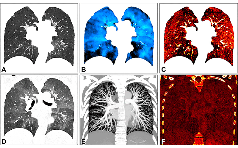
Images in a 34-year-old female patient show severe smoking-associated small airway disease with patchy ground-glass opacities and mosaic pattern. The images were generated with spectral postprocessing on a coronal plane at (A) inspiration (warped for voxelwise matching with expiration), (B) ventilation, (C) perfusion, (D) expiration, (E) CT angiography, and (F) late contrast enhancement. Quantitative parameters include whole lung or lobar volume, and inspiratory (A) and expiratory (D) attenuation and functional parameters such as ventilation (B), perfusion (C), and late contrast enhancement (F). They are described using descriptive statistics or histogram analysis. Corresponding functional maps show inhomogeneous ventilation with lobular air-trapping in the lower lobes and matched perfusion inhomogeneities. CT angiography (E) and late contrast enhancement (F) images show no abnormal findings.
https://pubs.rsna.org/doi/10.1148/radiol.230318 ©RSNA 2023
Other Promising Applications in Lung Imaging
The photon-counting CT protocol has other promising applications in lung imaging. It can provide important preoperative identification of areas of emphysema and perfusion defects in patients with chronic thromboembolic pulmonary hypertension, a progressive disease caused by blood clots that do not clear from the lungs.
Postoperatively, the protocol allowed evaluation of surgical success and was helpful in assessing the lungs after lung or stem cell transplant procedures. It may also be useful in follow-up of chronic obstructive pulmonary disease and looking at pathological findings in the lung tissue.
“We believe that the proposed protocol is generally valuable for diseases with known or unknown lung function impairment,” Dr. Shin said.
Dr. Shin and colleagues first applied the protocol to patients with interstitial lung disease. They then expanded the applications to include post-COVID-19 condition where interstitial lung disease sometimes develops.
“With the proposed protocol, we have also been able to answer many other questions related to post-COVID-19 condition, such as the detection of acute and chronic pulmonary emboli on CT angiography, and we are currently investigating whether perfusion changes can be quantified in microvascular damage or inflammatory areas,” Dr. Shin said.
The researchers are working to improve processing time and increase the robustness of the technique.
“Regional ventilation and perfusion depend on patient position and gravity, among other factors,” Dr. Shin said. “Further studies are needed to assess the dependence on position and depth of breathing, as well as the reproducibility of the measurements.”
For More Information
Access the Radiology study, “Regional Pulmonary Morphology and Function: Photon-counting CT Assessment.”
Read previous RSNA News articles about photon-counting CT: