‘Radiology’ Recognizes Top Images of 2022
Top images selected by Radiology in Training editors
Selections have been made for the 2022 Images in Radiology, an online medical imaging collection that is part of the journal Radiology. Each year, Images in Radiology publishes captivating images demonstrating important medical diagnoses and state-of-the-art medical imaging technology. These images exemplify the significant contributions made by radiology to the field of medicine.
Readership for Images in Radiology has grown significantly over the past several years leading to continuous growth in the number of eligible publications. For 2022, 43 images, published between July 2, 2021 and June 30, 2022, were eligible—up from 29 in 2021.
The top three images, second runner-up, first-runner-up and winner, were selected by the Radiology in Training editorial board based on aggregate scores from individual editorial board members’ rankings. The images were selected based on three criteria: novel technology or unusual pathology, educational or thought-provoking and visually compelling.
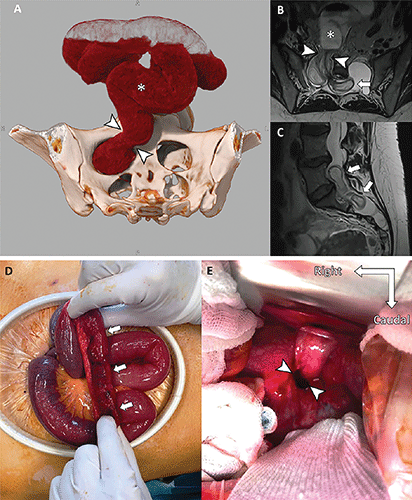
Images in 38-year-old woman with Marfan syndrome who was diagnosed with intradural small bowel herniation through a torn right S1 dural ectasia. (A) Oblique global illumination rendering of abdominal CT scan after semiautomatic segmentation of the enlarged afferent small bowel (*). The hernia orifice (arrowheads) separates the peritoneal cavity and the intradural space. (B, C) T2-weighted MRI scans in (B) axial and (C) sagittal planes show the herniated loop (arrowheads in B) of small bowel (* in B) ascending at L5-S1 (arrows), causing significant compression of the adjacent cauda equina nerve roots. (D) Intraoperative photograph of the incarcerated intestine with signs of distress (arrows). (E) Intraoperative photograph (anterior view) shows the hernia orifice in the sacrum (arrowheads). https://doi.org/10.1148/radiol.229031 ©RSNA 2022
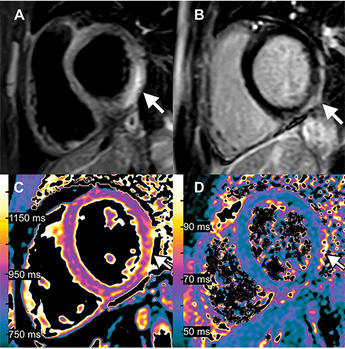
Images in 15-year-old boy with myocarditis after COVID-19 vaccination. One day after receiving his second vaccination dose, he developed fever, myalgia, and intermittent tachycardia. (A) T2-weighted short inversion time inversion recovery MRI scan at 1.5 T in short-axis view shows focal high-signal intensities (arrow) at basal lateral and inferior wall, indicating myocardial edema. (B) Late gadolinium enhancement image in short-axis view shows corresponding linear subepicardial enhancement (arrow), indicating inflammatory myocardial necrosis. (C) T1 mapping and (D) T2 mapping in short-axis view show elevated T1 and T2 at the mid ventricular lateral and inferolateral wall (arrow in C and D), indicating acute myocardial injury (focal T1, 1165 msec; focal T2, 70 msec; institution-specific cutoff values for acute myocarditis: T1global ≥1000 msec, T2 global ≥55.9 msec). https://doi.org/10.1148/radiol.229031 ©RSNA 2022
The first runner-up is an article entitled, “Myocarditis Following COVID-19 Vaccination,” by Alexander Isaak, Andreas Feisst and Julian A. Luetkens. 2022 marks the third consecutive year in which a COVID-related image was honored among top contenders for Images in Radiology, and this article attracted the most social media attention among all 2022 Images in Radiology receiving an Altmetric score placing it in the top 1% of all research outputs of the same age.
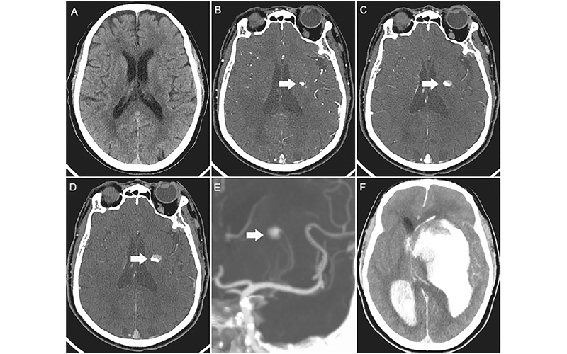
Images in a 73-year-old man with acute left-sided weakness. (A) Axial noncontrast CT image of the head shows no intracerebral hemorrhage. (B) Arterial phase CT angiogram obtained 192 seconds later, after administration of contrast material, demonstrates focal enhancement in the left basal ganglia (arrow). (C, D) CT angiograms obtained in the (C) second and (D) third phases, with delays of 39 seconds and 38 seconds, respectively, highlight the active expansion of the intracerebral hemorrhage with the blood-contrast level (arrows). (E) Coronal maximum intensity projection image reveals that the bleeding was located near the distal ends of the lenticulostriate arteries (arrow). (F) Follow-up noncontrast CT image obtained 1 hour after the third phase CT angiogram shows massive enlargement of the hematoma, intraventricular hemorrhage, hydrocephalus, and subfalcine herniation. https://doi.org/10.1148/radiol.229031 ©RSNA 2022
The second runner-up was a tie between two articles. The first, “CT of Ongoing Intracerebral Hemorrhage,” is by Chun Ma and Yi Zhou with imaging that captured the rapid evolution of a progressively expanding intracerebral hemorrhage in a 73-year-old man.
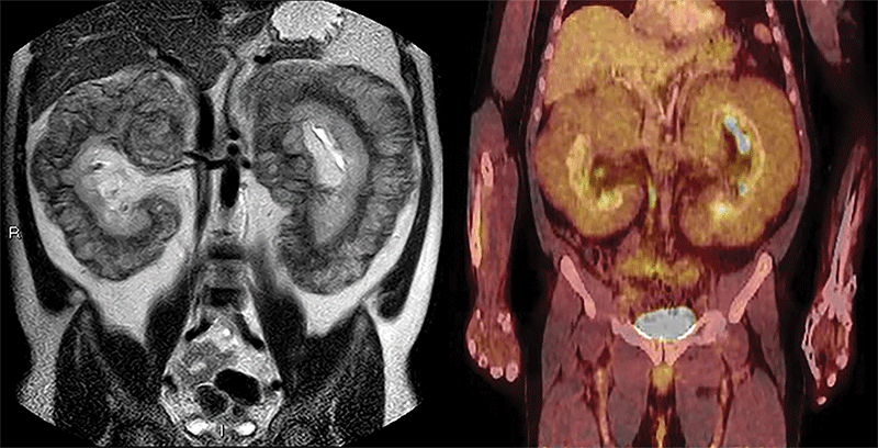
Images in a 47-year-old man with worsening lower extremity edema and chronic abdominal distention. Left: Coronal single-shot T2-weighted MRI scan shows massive bilateral perirenal infiltration by soft-tissue masses. Right: Fluorine 18 fluorodeoxyglucose PET/CT scan shows mild uptake by perirenal masses and upward displacement of liver. https://doi.org/10.1148/radiol.229031 ©RSNA 2022
The other second runner-up tie article is, “Massive Perirenal Involvement in Erdheim-Chester Disease,” by Jorge Polo-Sabau and Begoña López-Botet-Zulueta. The article includes an example of imaging of a rare disease. It serves as a reminder that even in the absence radiologic signs that reflect the most frequent appearances of certain disease conditions, radiologists may still consider certain diagnoses when a combination of imaging details and clinical presentation warrant it.
For More Information
Access the Radiology article, “2022 Top Images in Radiology: Radiology in Training Editors’ Choices.”
Review the 2021 Images in Radiology winners.