Dark-Field Chest Radiography Offers Potential Early Diagnosis of Pulmonary Emphysema
Interferometer into the beam path creates a dark-field image of emphysematous lungs


A dark-field chest radiography prototype developed by an interdisciplinary team at the Technical University of Munich, Germany, shows promise for early detection of pulmonary emphysema.
The current imaging modalities used for emphysema diagnosis—attenuation-based radiography and CT—do not detect early-stage disease, as they both rely on secondary signs at mid to late stages.
“Because the contrast in conventional radiography is based on the attenuation of the tissue, the surrounding tissues, such as the ribs and the heart are dominant in the images. If there are slight changes in the attenuation signal of the lung itself, it is very hard to see them,” said Theresa Urban, MSc, chair of biomedical physics at the Technical University of Munich and co-primary investigator of a Radiology study on this topic. “On conventional radiography, emphysema is only visible in later stages when there are secondary signs, such as a barrel shaped thorax and increased spacing between the ribs.”
Daniela Pfeiffer, MD, attending physician in the Department of Radiology at the Technical University of Munich School of Medicine, and co-senior author of the study, noted that even CT does not have the necessary resolution to detect and diagnose emphysema early on.
“The size of the alveoli is in the range of 50 to 150 micrometers. So even high-resolution CT is not able to visualize the changes at the level of the alveoli directly. CT shows us the destruction of lung parenchyma, but only in an advanced stage of the disease,” Dr. Pfeiffer said.
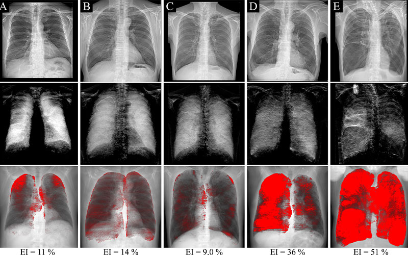
Attenuation-based (top row) and dark-field (middle row) radiographs and projections of CT-based emphysema quantification (bottom row) in five example participants with a significant emphysema index (EI) of 6% or more. The same window and level settings were applied within each respective modality. Signs of hyperinflation can be observed in attenuation-based images of participants with high emphysema severities. Areas of decreased dark-field signal correspond well to emphysematous areas on the CT-based emphysema projections. In B, the participant has a right-sided port catheter in place.
Dark-Field Provides Darker Patches on Lung Images
Whereas conventional radiography uses the attenuation of X-rays, dark-field radiography leverages the scattering of X-rays at material interfaces, such as the air-tissue interfaces in the lungs.
This is done with a standard radiography machine by inserting an interferometer with three gratings into the beam path. This allows the simultaneous capture of both a standard attenuation-based image and a dark-field image.
On a dark-field image, a healthy lung appears in uniform, bright white, due to intact alveolar structures that create a strong dark-field signal. The opposite occurs in a dark-field image of emphysematous lungs and is very apparent to the naked eye.
“In a healthy lung, the alveolar structure has many tissue interfaces, each of them causing a small change of direction in the beam that results in a strong scattering signal, or dark-field signal,” Urban said. “When there is some impairment of the alveolar structure, then there are fewer air-tissue interfaces, and therefore a weaker dark-field signal. In a dark-field image, this appears as dark patches on the lung.”
Dark-Field Chest Radiographs Has Potential As Diagnostic Tool
For the Radiology study, Urban, Dr. Pfeiffer and their team reviewed clinically indicated chest CTs of 83 patients with pulmonary emphysema.
The appearance of their lungs was also examined using both attenuation-based and dark-field radiography. Dark-field images were then compared to CT images to test any visual correlations of CT-based emphysema findings. This comparison showed that the locations of signal intensity loss on dark-field images corresponded to emphysematous areas found on CT.
“The emphysema detected in CT corresponds very well to the dark patches in the dark-field images. This tells us that the dark-field signal is accurate, and that it is sensitive to emphysematous changes of the lung,” said Urban.
To quantitatively measure the emphysema found in dark-field images, the researchers also calculated a dark-field coefficient using the dark-field signal and lung volume, generated per meter of tissue. Due to signal loss, this coefficient decreases in the presence of emphysema.
Although Dr. Pfeiffer notes that the coefficient is subject to testing with a larger sample size, it did appear to indicate the presence and severity of emphysema.
The dark-field coefficient was found to be negatively correlated with each patient’s CT-based emphysema index. Furthermore, participants with Fleischner Society grades of mild emphysema or higher exhibited a lower dark-field coefficient than those without emphysema.
In a Radiology editorial on this research, Hiroto Hatabu, MD, PhD, professor of radiology and Bruno Madore, PhD, associate professor of radiology, both at the Brigham and Women’s Hospital and Harvard Medical School in Boston, noted that the results of this study are significant because the use of CT for diagnosing emphysema has limitations. It is expensive, not readily accessible, and utilizes higher radiation doses. They posit that dark-field radiography might be an excellent diagnostic alternative in the future.
Dr. Pfeiffer emphasized the importance of diagnosing emphysema early, for which dark-field radiography may play a key role.
“Emphysema can't be cured or reversed to earlier stages, but treatments can slow its progression or relieve symptoms,” Dr. Pfeiffer said. “Therefore, it's crucial to diagnose early, and dark-field radiography may make that possible. With early therapy, we can significantly improve the outcomes for these patients.”
For More Information
Access the Radiology study, “Qualitative and Quantitative Assessment of Emphysema Using Dark-Field Chest Radiography,” and editorial, “Dark-Field Chest Radiography in the Detection of Emphysema."
Read previous RSNA News articles on emphysema and related topics: