Transgender Imaging Presents Unique Challenges for Radiology
Awareness of anatomic and pathologic changes can ensure accurate imaging interpretation

Read the full story in the May print issue of RSNA News.
There are an estimated 25 million transgender people worldwide, including 1.4 million who live in the U.S. They can have unique health care needs especially if they have undergone gender-affirming surgeries or are seeking preventive cancer screenings related to their sex assigned at birth or their identified gender.
Radiologists must be aware of the wide variety of anatomic and pathologic changes unique to patients who undergo gender-affirming surgeries to ensure accurate imaging interpretation. Unfortunately, detailed descriptions of imaging features remain sparse. There is also a lack of data related to cancer risk in transgender people.
Transgender people may be less likely to seek or receive necessary screenings due to a fear of discrimination, a lack of access to providers and dysphoria, or distress related to the mismatch between their gender identity and their sex assigned at birth.
“The radiologist is central to the care provided to transgender patients, not only in a consultative role for advising clinicians on the most appropriate imaging modalities, but also in the protocol selection, preparation of imaging technologists, and often in the performance of imaging procedures,” said Justin Stowell, MD, senior associate consultant, Department of Radiology, Mayo Clinic, Jacksonville, FL. “Moreover, a radiologist can serve as an advocate to ensure appropriate use of imaging for certain indications. Radiologists should be sensitive to these facts and make their imaging centers more welcoming, safe environments in which transgender patients may receive care.”
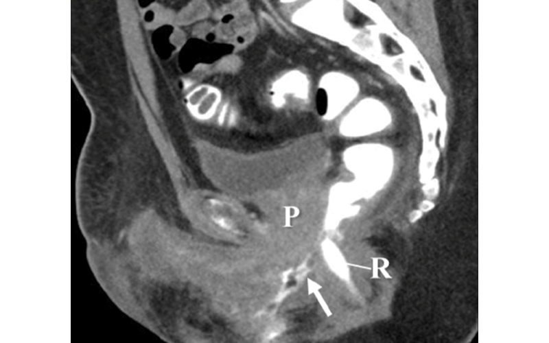
Dr. Stowell is one of the authors of the RadioGraphics study, "“Imaging Findings in Transgender Patients after Gender-affirming Surgery."
Challenges for Radiologists
Gender-affirming surgeries expand options for physical transition among transgender patients — those whose gender identity is incongruent with the sex assigned to them at birth. Growing medical insight, increasing public acceptance and expanding insurance coverage have improved access to and increased the demand for gender-affirming surgeries in the U.S. However, the numbers of gender-affirming surgeries remain low. In the 2015 U.S. Transgender Survey, only 25% of more than 27,000 respondents had undergone some form of gender-affirming surgery.
There has been a sizeable growth in the surgical literature about gender-affirming surgeries and their outcomes. However, detailed descriptions of the imaging features following these surgeries remain sparse.
“It is difficult to determine exactly why the radiology literature historically has remained so silent surrounding imaging of transgender people after gender-affirming surgery. Recently, there have been more publications in radiology journals describing expected post-operative imaging findings after gender affirming surgery perhaps as a result of increased general public awareness of the social and medical issues that affect transgender persons,” Dr. Stowell said. “However, more prospective, multi-institutional research is warranted.”
Another challenge for radiologists is the potential for error in misgendering — or providing inaccurate descriptions of imaging results — especially if the patient’s affirmed legal gender is reflected in the medical record.
“Radiologists should use caution when evaluating imaging studies in patients who have undergone gender-affirming surgery to avoid misgendering or assigning an inaccurate gender marker in reports. This may be more challenging if the patient is identified in the electronic medical record with their affirmed gender as opposed to that assigned to them at birth,” Dr. Stowell said.
For example, Dr. Stowell noted that a transmasculine person who has undergone hysterectomy and has affirmed, masculine gender markers and a legal name change, might have a radiologist who unknowingly assumes the patient should have male anatomic parts.
Therefore, changes after hysterectomy might be misinterpreted as the patient having a prostate if care is not taken during interpretation to assess the pertinent history. Conversely, the prostate is not removed during vaginoplasty in transfeminine patients, so a radiologist might misinterpret its presence as representing a mass.
Technology is recently allowing for the collection and display of more specific patient information and demographics, including gender identity, sex assigned at birth, chosen name and legal gender, according to Dr. Stowell, which can alert radiologists to important information about the patient, especially potential history of hormonal therapy and gender-affirming surgery.
For More Information
Access the study, “Imaging Findings in Transgender Patients after Gender-affirming Surgery." To claim CME credit for this activity, visit RSNA.org/Learning–Center.