Diffusion-Weighted MRI Accurate in Detecting Juvenile Idiopathic Arthritis of the Knee
Non-invasive, non-contrast method shows potential benefits for pediatric patients


Assessing arthritis in the knee can be difficult in adults using traditional MRI methods. In children — who may not be able to stay still or may even need to be sedated during the procedure — the process can be even more difficult.
A recent study published in Radiology, “Juvenile Idiopathic Arthritis: Diffusion-weighted MRI in the Assessment of Arthritis in the Knee,” by lead author Anouk Barendregt, MD, in the Department of Pediatric Immunology and Radiology at the Amsterdam University Medical Center, and colleagues, shows MRI technology may make the process of assessing arthritis easier for pediatric patients without sacrificing accuracy.
“We have been interested in novel methods to assess arthritis without injection of contrast agent for a long time,” Dr. Barendregt said. “This technique is much more patient-friendly, especially for children.”
Diffusion weighted imaging (DWI) is commonly used in areas such as stroke and prostate but has not been widely tested in the diagnosis and analysis of juvenile idiopathic arthritis. DWI has a number of benefits for pediatric patients, Dr. Barendregt said. It is non-invasive and there is no need to place an IV and administer contrast. The technique is also faster, so children don’t need to stay still as long, reducing the number of young patients that need sedation for a lengthy MRI scan.
Children with juvenile idiopathic arthritis often have autoimmune-mediated inflammation of the synovial membrane. The team studied the diagnostic accuracy of DWI for the detection of arthritis as compared with the clinical reference standard, which included contrast material–enhanced MRI.
The research team conducted a prospective study of 45 patients ages 6 to 18 who underwent pre- and post-contrast MRI of the knee with an additional DWI sequence between 2015 and 2018. For the clinical reference standard, a multidisciplinary team determined the presence or absence of arthritis on the basis of clinical, laboratory and imaging findings, excluding DWI. The two data sets were then scored by two radiologists blinded to all clinical data.
DWI Shows High Accuracy
The study found that DWI was accurate in detecting arthritis in pediatric patients, both in sensitivity and specificity. The results showed sensitivity for detection of arthritis with DWI was 93% (13 of the 14 participants with arthritis were correctly classified). DWI also scored above 80% on specificity for patients without arthritis. The results indicate that DWI could replace contrast-enhanced MRI for imaging of inflammation.
“What we found in our study quite surprised us — it was better than we expected,” said study co-author Robert Hemke, MD, PhD, musculoskeletal radiologist at Amsterdam University Medical Center, who said the team has been researching DWI in arthritis patients for about five years. “During this time, we had regarded contrast-enhanced MRI as the reference standard.”
The Radiology study, which began as a small research project several years ago, was aided significantly by the participation of pediatric rheumatologists and other researchers.
“Working as a team really benefited our research,” Dr. Hemke said. “I’ve learned how important it is to keep your eyes open to other fields of imaging outside of the musculoskeletal domain.” With further and larger validation studies, DWI may be valuable for both adult and pediatric arthritis patients, researchers said.
For More Information
Access the Radiology study, “Juvenile Idiopathic Arthritis: Diffusion-weighted MRI in the Assessment of Arthritis in the Knee.”
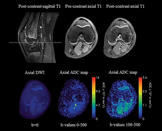
Image in 12-year-old girl without synovial inflammation in whom a functional disorder and/or pain syndrome was eventually diagnosed. The score from the diffusion-weighted imaging (DWI) data set was in agreement with the reference standard: All members of the multidisciplinary team agreed that no arthritis was present in the knee. ADC = apparent diffusion coefficient. Arrows indicate areas of synovial inflammation. ADC = apparent diffusion coefficient.