Zero Echo-Time MRI Shows Diagnostic Efficacy Similar to CT
Method shows potential to become the preferred radiation-free modality for pediatric bone imaging

Children in the U.S. undergo an estimated five to nine million diagnostic CT examinations per year. Because pediatric patients are more vulnerable to the potential effects of radiation, programs to reduce radiation dose in children have gained traction over time.
While clinicians endeavor to adhere to Image Gently® principles, researchers are also investigating alternative imaging options. Currently, CT is still required for important diagnostic indications, including those related to acute screening and cortical bone imaging.
In her 2018 Siemens Healthineers/RSNA Research Scholar grant project, Mai-Lan Ho, MD, worked with colleagues to evaluate the use of zero echo-time (ZTE) MRI to achieve radiation-free pediatric bone imaging.
“ZTE MRI is a promising alternative modality to CT that uses ultrafast readouts to visualize cortical bone in relatively short examination times,” she said.
ZTE MRI is a technique that produces images similar to those obtained with X-ray or CT. It utilizes ultrafast readouts to capture signal from short-T2 tissues. Dr. Ho is professor of radiology and director of the translational imaging and advanced neuroimaging cores at The Ohio State University and Nationwide Children’s Hospital in Columbus. She also serves as faculty lead for the institution’s imaging genomics research affinity group, chair of the Asian Pacific American Network and secretary for the Association of Staff and Faculty Women.
“With this project, we proposed to develop and translate ZTE MRI for practical clinical use, focusing on inclusion of diverse populations and pathologies in neuroradiology, head and neck, and pediatric imaging,” Dr. Ho said. “These fields present major technical challenges, but also offer great potential for scientific advancement.”
Project Aims Focus on Clinical Workflow, Diagnostic and Treatment Efficacy
For the study, Dr. Ho and colleagues first focused on establishing a standard clinical ZTE workflow. Over the course of a few years, the team successfully optimized ZTE at two different institutions for a variety of MRI platforms and clinical indications, accruing a total of over 250 subjects.
“We also experimented with different image processing approaches to generate synthetic CT or ‘bright-bone’ images from raw ‘black-bone’ ZTE data,” she said.
According to Dr. Ho, the group’s second aim was to compare the efficacy of both ZTE and CT for patient diagnosis and interventional planning. She noted that their efforts were successful as the two techniques are qualitatively and quantitatively comparable, though ZTE requires specialized downstream processing and user training.
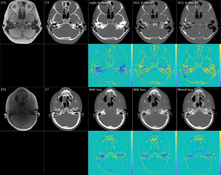
Comparison of ZTE, ground truth CT, and deep learning-generated synthetic CT [top rows] with color difference maps [bottom rows] for various neural networks and loss functions. Courtesy of Sven Bambach, PhD.
Experience Highlights Inherent “Bench-to-Bedside” Challenges
Among the challenges of the study was a familiar problem: the COVID-19 pandemic.
“During COVID-19, we explored flexible enrollment pathways for potential study subjects and referring physicians, while also utilizing deep learning strategies to analyze our existing data,” Dr. Ho said. “Multicenter collaborations were extremely useful for stimulating interest and promoting operationalization.”
Beyond pandemic-related issues, Dr. Ho and her team spent considerable time and energy deploying ZTE at multiple institutions for different MRI platforms and pulse sequences, an effort that involved several physicist and vendor partnerships.
Throughout the planning stages and while conducting the study, Dr. Ho said she learned from the diverse viewpoints of all stakeholders involved.
“I was fascinated by the range of attitudes regarding potentially disruptive technologies represented by advanced imaging and artificial intelligence,” she said. “Proposed workflows and use cases, where the algorithm could potentially be used. varied widely across institutions and individuals, and questions arose during patient scanning, image reconstruction, radiologist reporting and downstream data processing.”
Despite the apparent safety and practicality of the technology, Dr. Ho acknowledged that additional work is necessary to increase public awareness and education, build physician understanding and adoption, and optimize technical improvements to target specific disease applications.
“Overall, my experience highlights the challenges inherent in the translational ‘bench-to-bedside’ imaging pipeline,” she said.
Continuing her efforts to improve care for the most vulnerable patients, Dr. Ho has focused on implementing techniques like ZTE to enable better, faster and safer imaging for children.
“Being cross-trained in both adult and pediatric neuroradiology, I hope that integration within and among specialties will provide greater continuity of care across the lifespan,” Dr. Ho said. “Bringing more institutions on board will help generate more diverse training data for AI algorithms and mobilize comparative effectiveness studies to spur widespread adoption of new technologies.”
“Receiving the Research Scholar Grant at a critical early stage of my career empowered me to secure a research faculty position with dedicated mentorship and protected time. This gave me an opportunity to transition from my initial focus on clinical excellence while maintaining strong relationships with referring physicians.”
Mai-Lan Ho, MD
R&E Funding Opens New Pathways of Learning, Leadership
“Receiving the Research Scholar Grant at a critical early stage of my career empowered me to secure a research faculty position with dedicated mentorship and protected time,” Dr. Ho said. “This gave me an opportunity to transition from my initial focus on clinical excellence while maintaining strong relationships with referring physicians.”
The dedicated research time enabled her to acquire new technical knowledge, improving her skills in advanced imaging and AI while working alongside imaging physicists and data scientists.
Dr. Ho attributed several positive outcomes to her grant funding, including the development of her unique reputation in advanced pediatric neuroimaging and precision health. “I leveraged these successes into multiple scientific publications and technical reports, as well as federal, foundation and industry grants,” she said.
For More Information
Learn more about R&E Foundation grants at RSNA.org/Research/Funding-Opportunities.
Read previous RSNA News stories about R&E Foundation grants: