Bladder Cancers In The Ureteral Orifice Have Higher Frequency Of Detrusor Muscle Invasion
VI-RADS can be used to predict muscle-invasive bladder cancer


Bladder cancer is the most frequent urinary system malignancy and it is associated with a high recurrence rate.
Treatment for bladder cancer is dependent on the status of muscle invasion. Non-muscle-invasive bladder cancer (NMIBCa) is usually low grade, while muscle-invasive bladder cancer (MIBCa) is an aggressive tumor with a poor prognosis and requires more intensive treatment.
Imaging is required to provide precise preoperative characterization of the muscle invasion. Recent developments in MRI have made multiparametric MRI an important complement to transurethral resection of the bladder tumor.
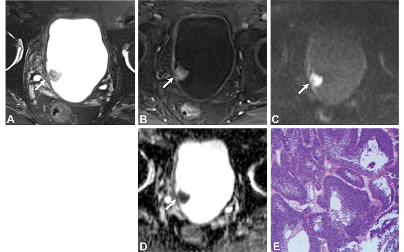
The Vesical Imaging Reporting and Data System (VI-RADS) offers a five-point MRI score to predict the likelihood of muscle infiltration by bladder cancer. However, the VI-RADS criteria were reported to have limitations, particularly for tumors located at ureteral orifices.
“Numerous studies have shown that VI-RADS is an effective comprehensive tool in the detection of muscle invasion for bladder cancer,” according to Huanjun Wang, MD, PhD, from the Department of Radiology, The First Affiliated Hospital, Sun Yat-Sen University, in Guangzhou, China. “We wanted to look at whether VI-RADS could be used to accurately predict muscle invasion for tumors occurring at the ureteral orifice, a specific anatomic location with a higher frequency of detrusor muscle invasion by tumor.”
https://doi.org/10.1148/radiol.220028 © RSNA 2022
Radiologists Were Able to Match VI-RADS Staging
In this retrospective study, patients with histopathologically confirmed bladder cancer occurring at the ureteral orifice were analyzed. Two blinded radiologists independently scored multiparametric MRI scans according to VI-RADS.
A total of 78 patients were included in the final analysis: 25 with NMIBCa and 53 with MIBCa. At consensus reading, one case (1%) was scored as VI-RADS 1, 27 cases (35%) were scored as VI-RADS 2, six (8%) were scored as VI-RADS 3, 10 (13%) were scored as VI-RADS 4, and 34 (44%) were scored as VI-RADS 5.
On comparison of the VI-RADS score with histopathologic findings, it was confirmed that the presence of muscle invasion was 0% (zero of one) for VI-RADS 1, 15% (four of 27) for VI-RADS 2, 83% (five of six) for VI-RADS 3, 100% (10 of 10) for VI-RADS 4, and 100% (34 of 34) for VI-RADS 5. The area under the receiver operating characteristic curve (AUC) of VI-RADS in the detection of MIBCa was 0.96, indicating a high diagnostic test accuracy.
“At present, bladder cancer staging is accomplished by the combination of examination, transurethral resection of bladder tumor specimens and imaging. However, the quality of the transurethral resection of bladder tumors often varies among surgeons and they may miss muscle infiltration in up to 25% of invasive cancers. Therefore, there is an urgent need to develop an accurate and noninvasive method to assess muscularis invasion in bladder cancer,” said co-author, Yan Guo, MD, PhD, also from Sun Yat-Sen University. “The area under the receiver operating characteristic curve of VI-RADS in the detection of MIBCa was high, suggesting that VI-RADS can accurately distinguish non-muscle-invasive and muscle-invasive bladder cancer prior to surgery and provide a powerful tool for patients to choose treatment modalities.”
Accurately determining the muscle invasion status of bladder cancer prior to surgery is critical due to the differences in available treatment options. Radiologists therefore play an important role in preoperative imaging, according to Qian Cai, MD, co-author from Sun Yat-Sen University.
“It’s essential to know whether or not the cancer is invasive of muscle or not due to the severity of the treatments,” Dr. Cai said. “Muscle-invasive bladder cancer must be treated with radical cystectomy, whereas non-muscle-invasive bladder cancer treatment is less aggressive. The high complication rate and low quality of life following the radical cystectomy surgery makes it essential to determine the muscle invasion status of bladder cancer prior to surgery.”
Further evaluation is needed to continue studying bladder cancers located at the ureteral orifice.
“VI-RADS scoring, specifically the cutoff of 1–2 versus the cutoff of 3–5, could be used to accurately predict muscle-invasive bladder cancer. Further evaluation of this scoring system is warranted,” Dr. Wang said.
For More Information
Access the Radiology study, “Multiparametric MRI Evaluation of VI-RADS for Bladder Tumors Located at the Ureteral Orifice.”
Read previous RSNA News articles about genitourinary imaging: