AI Helps Detect Brain Aneurysms on CT Angiography
Deep learning a tool to support interpretation of cerebral aneurysms

Deep learning (DL) can help physicians detect potentially life-threatening cerebral aneurysms on CT angiography, according to a study published in Radiology.
CT angiography is usually the first choice for evaluating cerebral aneurysms, but cerebral aneurysms can be overlooked on the initial assessment due to their small size and the complexity of the blood vessels in the brain.
“In our daily work we are always faced with cases in which some important lesions have been missed by the human eye,” said study senior author Xi Long, PhD, from the Department of Radiology at Tongji Medical College’s Union Hospital in Wuhan, China. “Cerebral aneurysms are among those small lesions that may be overlooked on the routine assessment of radiological images.”
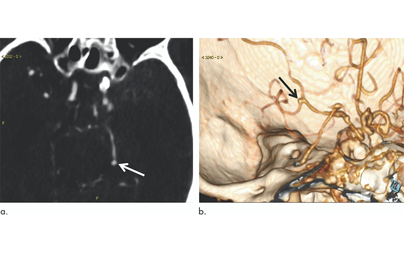
DL Can Enhance Radiologists’ Image Interpretation
In the new study, Dr. Long and colleagues developed a fully automated, highly sensitive algorithm for the detection of cerebral aneurysms on CT angiography images. They used CT angiograms from more than 500 patients to train the DL system, and then they tested it on another 534 CT angiograms that included 649 aneurysms.
The algorithm detected 633 of the 649 cerebral aneurysms for a sensitivity of 97.5%. It also found eight new aneurysms that were overlooked on the initial assessment.
Statistical analysis revealed that DL assistance enhanced radiologists’ performance. The improvement was most pronounced in the less experienced radiologists.
“The developed deep learning system has shown excellent performance in detecting aneurysms,” Dr. Long said. “We found some aneurysms that were overlooked by the human readers on the initial reports, but they were successfully depicted by the deep learning system.”
The results suggest that the DL algorithm has promise as a supportive tool for detecting cerebral aneurysms with a potential to be used clinically for a second opinion during interpretation of head CT angiography images. It has a number of advantages in this setting, Dr. Long said, primarily due to the fact that the computer is not influenced by factors like the level of experience, working time and mood that affect human performance.
The system has some limitations, Dr. Long noted. It can miss very small aneurysms or aneurysms located close to similar density structures like bones. It also suffers from false positive results, which necessitates careful revision of the system suggestions by human readers.
“Simply put, the deep learning system is intended to assist human readers, not to replace them,” Dr. Long said.
The system needs further validation on more heterogeneous data, such as images from people in different parts of the world, which is essential for assessing its generalizability and applicability to daily clinical work.
“At this time, the role of this deep learning system, which has been trained to recognize aneurysms, is to give suggestions to the human reader to improve their performance and reduce mistakes,” Dr. Long said. “The combined work of the human reader and computer system improves the diagnostic accuracy for the patient’s sake.”
For More Information
Access the Radiology study, “Deep Learning for Detecting Cerebral Aneurysms with CT Angiography.”
Read previous Radiology study stories:
- Deep Learning Model Provides Rapid Detection of Stroke-causing Blockages
- Vertebral Augmentation May Improve Mortality in Patients with Osteoporotic Fractures