Advances in Image-Guided Percutaneous Ablation Techniques Aid Cancer Treatment
Augmented reality and irreversible electroporation show promise as ablation techniques


An excerpted version of this article appears below. Read the full article in the March print issue of RSNA News and access the Radiology study linked below.
As the trend continues toward non-invasive medical procedures, image-guided percutaneous ablation is becoming increasingly popular as an effective alternative to surgery for the treatment of tumors in the liver and other organs. And although image-guided percutaneous ablation technology still has a number of drawbacks, the evolution of techniques and applications is taking the technology in promising new directions.
Researchers at the Cleveland Clinic have had success using augmented reality (AR) in mitigating one of the risks of ablation — damage to surrounding healthy tissue when targeting tumors. “A key part of an ablation procedure is critical structural avoidance,” said Charles Martin, MD, an interventional radiologist at the Cleveland Clinic, who presented a session on this topic at RSNA 2019. “Radiologists can become so fixated on what we need to target that we sometimes forget about navigating around the things we want to avoid. Augmented reality can help us do both.”
The technique being developed at the Cleveland Clinic superimposes digital twins of interventional tools and patient-specific anatomies in the form of 3D holograms. Unique algorithms spatially and anatomically register these holograms to the patient. Using a head-mounted display, clinicians have “X-ray vision” into the patient’s anatomy and can see the position of the surgical instrument with respect to these structures.
Furthermore, an electromagnetic (EM) field is generated around the patient and tracks the operative tools in relation to the 3D CT hologram. The EM displays a light-ray from the needle, thereby showing the radiologist the trajectory of the instrument and how it will intersect with internal anatomical structures.
Finally, the system fuses real-time ultrasound (US) images with the registered CT, letting the radiologist see the US image displayed anatomically within the patient as they scan over their skin, creating a sort of flashlight into the patient’s body. This verifies the registration of the CT and allows for a more accurate probe placement relative to the preoperative plan.
“Pre-procedurally, this coupling of AR and holography with electromagnetic guidance allows us to improve the speed of tumor localization,” Dr. Martin said. “It can also give radiologists confidence when placing the probes interprocedurally, meaning more accurate ablations with fewer complications.”
A Non-Thermal Ablative Technique for Destroying Pancreatic Tumors
Historically, the risk of causing thermal-related injuries to crucial blood vessels, bile ducts and gastrointestinal structures has made percutaneous ablation of limited use to treating pancreatic cancer. But considering that pancreatic cancer is the fourth leading cause of cancer death in the U.S. (with a five-year relative survival rate of less than 8%), researchers have been searching for a suitable alternative. One candidate is irreversible electroporation (IRE) – the subject of a recent Radiology study.
“IRE is a primarily nonthermal ablative technique in which high-voltage electrical pulses are applied between needle electrodes inserted within and around the tumor,” said Martijn R. Meijerink, MD, PhD, a radiologist at Amsterdam University Medical Center and the study’s lead author. “The pulses irreversibly damage the cellular membrane by creating nanopores, inducing programmed cell death.”
While other studies — mostly retrospective — have confirmed the safety profile of IRE, most research has had limited sample sizes and a relatively short follow-up. The aim of the Radiology study was to critically assess the efficacy and long-term safety of percutaneous IRE for locally advanced pancreatic cancer and isolated local recurrence following surgical resection of pancreatic cancer.
Between December 2012 and September 2017, 50 patients, 40 with locally advanced pancreatic cancer and 10 with local recurrence after resection, were treated with percutaneous IRE. The target median OS was 11.6 months for participants receiving no induction chemotherapy or gemcitabine-based induction chemotherapy and 14.9 months for those receiving induction of FOLFIRINOX (5-fluorouracil, leucovorin, irinotecan, and oxaliplatin).
“The primary results of our study were the median survival times in participants with primary locally advanced pancreatic cancer increased to 17 months and local recurrence to 16 months, both exceeding the target median survival times based on chemotherapy alone,” Dr. Meijerink said. “These findings suggest a survival benefit compared with current standard of care.”
For More Information
Access the Radiology study, “Percutaneous Irreversible Electroporation in Locally Advanced and Recurrent Pancreatic Cancer (PANFIRE-2): A Multicenter, Prospective, Single-Arm, Phase II Study.”
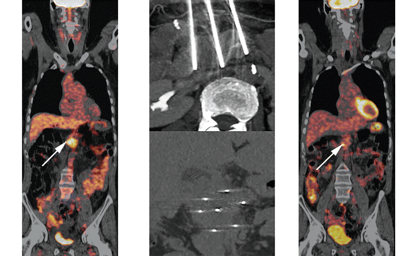
A, PET/CT showed an FDG-avid pancreatic tumor (arrow) in a 49-year-old that was successfully eradicated. B, no tracer uptake (arrow) was seen 18 months after IRE. C, During IRE, three needles surrounded the tumor (axiel view) and seven needles covered the entire tumor (coronal view). Ruarus AH, etal, Radiology 2020 ©RSNA 2020