AI Improves Efficiency and Accuracy of Digital Breast Tomosynthesis
Study shows increase in sensitivity and specificity, decrease in reading time

Artificial intelligence (AI) helps improve the efficiency and accuracy of an advanced imaging technology used to screen for breast cancer, according to a new study published in Radiology: Artificial Intelligence.
Research has shown that digital breast tomosynthesis (DBT) improves cancer detection and reduces false-positive recalls compared to screening with digital mammography (DM) alone. However, the DBT exam can take almost twice as long to interpret as DM due to the time it takes for the radiologist to scroll through all the images. This increased time is likely to be more consequential as DBT increasingly becomes the standard-of-care for mammographic imaging.
For the study, researchers developed a deep learning (DL) system to identify suspicious findings in the DBT images. After developing and training the system, the researchers tested its performance by having 24 radiologists, including 13 breast subspecialists, each read 260 DBT examinations with and without AI assistance. The examinations included 65 cancer cases.
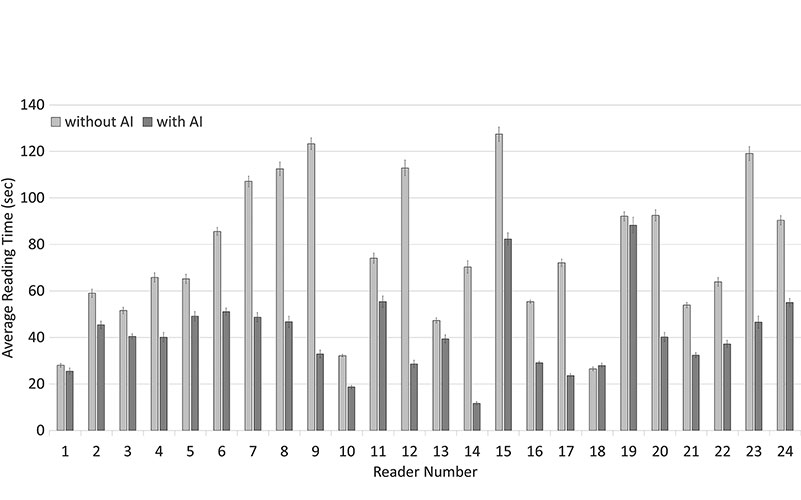
AI Helps Radiologists Shorten Image Reading Time
Use of AI was associated with improved accuracy and shorter reading times. Sensitivity increased from 77% without AI to 85% with it. Specificity increased from 62.7% without AI to 69.6% with it. The recall rate for non-cancers decreased from 38% without AI to just 30.9% with it. On average, reading time decreased from just over 64 seconds without AI to only 30.4 seconds with it.
“Overall, readers were able to increase their sensitivity by 8 percent, lower their recall rate by 7 percent and cut their reading time in half when using AI concurrently while reading DBT cases compared to reading without using AI,” said study lead author Emily F. Conant, MD, professor and chief of breast imaging from the Department of Radiology at the Perelman School of Medicine at the University of Pennsylvania in Philadelphia.
Also showing improvement was the area under the receiver operating characteristic curve (AUC). Radiologist performance increased from 0.795 without AI to 0.852 with AI.
“We know that DBT imaging increases cancer detection and lowers recall rate when added to 2D mammography and even further improvement in these key metrics is clinically very important,” Dr. Conant said. “And, since adding DBT to the 2D mammogram approximately doubles radiologist reading time, the concurrent use of AI with DBT increases cancer detection and may bring reading times back to about the time it takes to read DM-alone exams.”
The researchers expect the DL approach to improve as it is exposed to larger and larger data sets, making its potential impact on patient care even more significant.
“The results of this study suggest that both improved efficiency and accuracy could be achieved in clinical practice using an effective AI system,” Dr. Conant said.
For More Information
Access the Radiology: Artificial Intelligence study, “Improving Accuracy and Efficiency with Concurrent Use of Artificial Intelligence for Digital Breast Tomosynthesis” and read the accompanying editorial, "Using Time as a Measure of Impact for AI Systems: Implications in Breast Screening."