Oscillating Microbubbles Could Revolutionize Ultrasound
Microbubbles — which were initially created as vascular contrast agents for ultrasound (US) imaging — are now taking US in promising new directions in cancer treatment and other areas of medicine, according to a leading expert in the field.
The U.S. Food and Drug Administration (FDA) first approved microbubbles — gas-filled bubbles about the size of a human red blood cell — for use with US in the early 2000s. These injectable bubbles were initially used in cardiology, but the number of applications has increased in recent years as researchers have begun to exploit their ability to oscillate, or grow and collapse. This oscillation, under the precise control of acoustic pressure from the transducer, has opened new venues in both diagnosis and therapy for cancer and other diseases.
For example, microbubbles, when paired with subharmonic imaging technology, can be used as pressure sensors, a discovery that led the way to the development of a noninvasive pressure measurement technique known as subharmonic-aided pressure estimation (SHAPE), explained Dr. Forsberg, who delivered the 2018 RSNA New Horizons lecture on this topic.
“These pressure estimates represent something that is fundamentally different and that no other imaging mode can do right now,” Dr. Forsberg said.
SHAPE measurements are a potential noninvasive alternative to catheter-based measurements of pressure in the heart — measurements that provide important information for heart transplant patients and those with cardiovascular disease. Costs and use of ionizing radiation are the chief drawbacks of the invasive approach.
A 2017 proof-of-concept study on 15 patients led by Dr. Forsberg’s Thomas Jefferson University colleague Jaydev Dave, PhD, showed that pressure errors between the subharmonic technique and the catheter were very low (<3.5 mmHg), lending support to SHAPE’s viability for noninvasive pressure measurements.
“These numbers are so good that even cardiologists are paying attention,” said Dr. Forsberg. “We are now running a larger clinical trial of about 130 patients. We think if we continue achieving this level of success we will be able to do something you can’t do with any other imaging modality.”
Potential in Hypertension, Breast Cancer
In a 2013 Radiology study of 45 patients, SHAPE showed good overall agreement with catheter-based measurements for estimating portal hypertension, especially in patients at increased risk for variceal hemorrhage, a potentially life-threatening effect of the condition. Results of the larger clinical trial also look promising.
“Based on about 135 patients, we have seen 90 to 94 percent accuracy for noninvasively diagnosing portal vein hypertension,” Dr. Forsberg said.
The use of oscillating microbubbles as pressure sensors could also provide early indications of the effectiveness of chemotherapy in treating breast cancer. The approach takes advantage of the fact that interstitial fluid pressure is significantly higher in a tumor than in normal tissue.
A 2017 study in Radiology found that SHAPE was able to predict treatment response in breast cancer patients with 100 percent accuracy after just one round of chemotherapy. (See Web Extras.)
“We see this as an indication that the pressures inside the tumor have dropped, these vessels have opened up and you’re getting more signal in there,” Dr. Forsberg said. “This is an indication that chemotherapy is working.”
Microbubbles might also improve radiation therapy by acting as vehicles for oxygen delivery. Hypoxia, or lack of oxygen, makes tumors resistant to the effects of radiation therapy, so researchers tried delivering oxygen-filled microbubbles to the site of a tumor and then destroying them, releasing the oxygen into the tumor.
In a study of 50 mice, the combination of oxygen delivery and radiation therapy significantly increased tumor volume control.
Because the FDA has not yet approved this approach in humans, researchers are studying US-targeted microbubble destruction (UTMD) as a more immediate option.
Therapeutic Approaches with Microbubbles
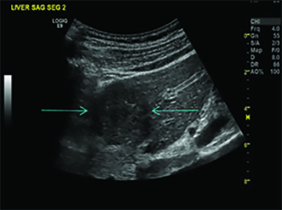
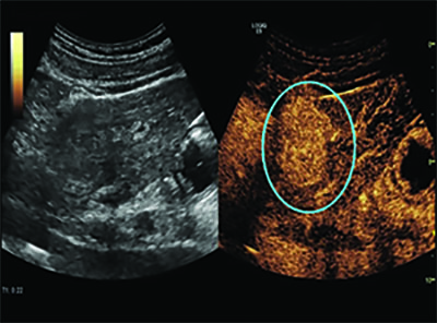
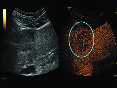
Dr. Forsberg’s colleague, John Eisenbrey, PhD, is in the early phase of a clinical trial studying UTMD on liver cancer patients undergoing radioembolization. The researchers introduced the microbubbles into the tumors and then destroyed them with acoustic waves, making the tumors more sensitive to the radiation. Follow-up imaging showed that of the seven patients who underwent UTMD, two experienced complete response to treatment and four had partial response. In contrast, only one of the four patients who did not undergo UTMD had a partial response and three had no response at all.
“We are about a quarter of the way into our ongoing clinical trial, but we think these preliminary efficacy results show us that there is potential for this to work,” Dr. Forsberg said.
In addition, researchers are studying oscillating microbubbles as a way to deliver drugs to the brain through the almost impermeable blood-brain barrier (BBB).
Research out of the University of Toronto on five Alzheimer’s disease patients showed that oscillating microbubbles could be used to safely, reversibly and repeatedly open the barrier. The researchers now want to attempt drug delivery through the temporary BBB opening.
Microbubbles also appear to improve the uptake of chemotherapeutic drugs in tumors. Using a technique called sonoporation, Norwegian researchers applied US to induce microbubbles vibrations making blood vessels in pancreatic cancer more permeable to chemotherapy drugs.
The 10 patients were able to tolerate more treatments, and median survival nearly doubled, from nine months to 17 months, without increased chemotherapy dosage and with no added toxicity or additional side effects.
The study, led by Odd Helge Gilja, MD, of the University of Bergen, was published in 2013 as well as 2016 and a larger trial is ahead, according to Dr. Forsberg.
WEB EXTRASAccess the Radiology study, “Monitoring Neoadjuvant Chemotherapy for Breast Cancer by Using Three-dimensional Subharmonic Aided Pressure Estimation and Imaging with US Contrast Agents: Preliminary Experience,” at RSNA.org/Radiology.