
RSNA Press Release
RSNA 2026 Margulis Award Goes to Deepfake Medical Imaging Study
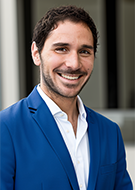
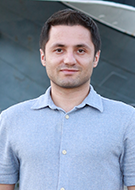
OAK BROOK, Ill. (Oct. 20, 2026) – Research into how AI-generated X-ray images may fool radiologists and large language models (LLMs) has earned the 2026 Alexander R. Margulis Award for best original scientific article published in Radiology, the flagship journal of the Radiological Society of North America (RSNA).
“The study captured the zeitgeist of a crucial moment in medicine, where new AI capabilities raise fundamental questions about authenticity of and trust in medical imaging,” said Suhny Abbara, M.D., editor of Radiology and editor-in-chief of RSNA’s journal portfolio. “By systematically evaluating the ability of radiologists and AI tools to identify AI-generated radiographs, the authors moved the conversation from speculation to evidence. The findings have received widespread attention and sparked a thoughtful ongoing debate about emerging risks and the potential need to establish safeguards.”
Generative AI tools can now create realistic medical images from plain-language prompts. These high-fidelity synthetic images, commonly known as deepfakes, can be difficult to distinguish from authentic ones.
While they may help expand rare-disease datasets, train algorithms and enrich radiology education, their growing realism and accessibility also raise concerns about trust and security.
“The release of increasingly accessible image-generation tools such as ChatGPT Image in 2025 made it possible for almost anyone to create highly realistic synthetic medical images,” said the study’s co-lead author, Mickael Tordjman, M.D., M.S., a postdoctoral fellow at the Biomedical Engineering and Imaging Institute at the Icahn School of Medicine at Mount Sinai in New York City. “We wanted to proactively test whether human experts and state-of-the-art multimodal LLMs could distinguish these synthetic radiographs from authentic ones, in order to highlight both the opportunities and the vulnerabilities related to this technology.”
The three-phase retrospective study included 17 radiologists from 12 centers across six countries. The readers represented several subspecialties and ranged from senior residents to radiologists with up to 40 years of experience.
The researchers assembled two balanced image sets. The first included 154 X-rays from multiple anatomic regions, containing 77 authentic images and 77 synthetic images generated using GPT-4o.
The second set included 110 chest X-rays, containing 55 authentic images and 55 synthetic images generated using the radiology-specific RoentGen model.
In the first phase, the radiologists were not told the purpose of the study. They evaluated image quality, noted artifacts and provided diagnoses. Only seven of 17 readers (41%) spontaneously reported that AI-generated images were present. Diagnostic accuracy for abnormalities was similar for the authentic X-rays (91.3%) and the synthetic X-rays (92.4%).
In phase two, after being told some images were synthetic, the radiologists classified each image as authentic or AI-generated. Their mean accuracy was 74.8% for the GPT-4o dataset and 70% for the RoentGen dataset.
“The most surprising finding was that even when radiologists were explicitly told that synthetic images were present in the dataset, their mean accuracy was moderate (approximately 75%), and years of professional experience offered no advantage in recognizing synthetic images,” said co-lead author Murat Yuce, M.D., M.S., a postdoctoral fellow also at Mount Sinai. “The fact that highly trained radiologists are not able to recognize all the synthetic images suggests that generative imaging technology has already reached a remarkable level of realism.”
In the final phase, four multimodal LLMs, GPT-4o, GPT-5, Gemini 2.5 Pro and Llama 4 Maverick, also performed the classification task. No model detected every synthetic X-ray. For the GPT-4o-generated images, GPT-4o achieved 85.1% accuracy and GPT-5 achieved 82.5%, compared with 56.5% for Gemini 2.5 Pro and 59.1% for Llama 4 Maverick.
The findings suggest that visual inspection alone may not be enough to verify whether a medical image is authentic.Dr. Tordjman suggested that health systems could integrate automated cryptographic authentication and additional safeguards into imaging workflows, but the concern extends beyond clinical care.
“There is also a risk that synthetic medical images could be presented as authentic on social media or even in scientific publications, making traceability increasingly important,” he cautioned.
According to Dr. Yuce, transparency is essential for responsible use.
“All synthetic images should be clearly identified as such and never presented as genuine,” he said. “At the same time, genuine radiologic images should also be protected through mechanisms that help verify their authenticity.”
In the near term, Dr. Yuce noted that practical approaches such as watermarking, cryptographic hashing or embedding imperceptible pixel-level information—without affecting diagnostic quality—may offer relatively expedient and reliable safeguards.
Looking ahead, more sophisticated methods, such as AI-based detection systems, could serve as medium- to long-term solutions. Education will also be important.
“Radiology training programs could integrate synthetic imaging to demonstrate its advantages and potential risk,” Dr. Yuce said. “The field can capitalize on the educational advantages of generative AI while safeguarding clinical integrity.”
The Alexander R. Margulis Award for Scientific Excellence recognizes the best original scientific article published in Radiology between July 1 of the prior year and June 30 of the award year. The main criteria for selection are scientific quality and originality. Selections are made by the Nominating Committee and the Selection Committee.
The award is given each year in honor of the late Alexander R. Margulis, M.D., a distinguished investigator and inspiring visionary in the science of radiology and longtime chair of the Department of Radiology at the University of California, San Francisco.
The Margulis Award will be presented to Drs. Tordjman and Yuce during RSNA 2026 at McCormick Place in Chicago at the inaugural Radiology Best Science session on Sunday, Nov. 29 at 1 p.m. CT
Access the Radiology study, “The Rise of Deepfake Medical Imaging: Radiologists’ Diagnostic Accuracy in Detecting ChatGPT-generated Radiographs.”Radiology is edited by Suhny Abbara, M.D., FACR, MSCCT, Mayo Clinic, Jacksonville, Florida, and owned and published by the Radiological Society of North America, Inc. (https://pubs.rsna.org/journal/radiology)
RSNA is an association of radiologists, radiation oncologists, medical physicists and related scientists promoting excellence in patient care and healthcare delivery through education, research and technologic innovation. The Society is based in Oak Brook, Illinois. (RSNA.org)
Images (JPG, TIF):
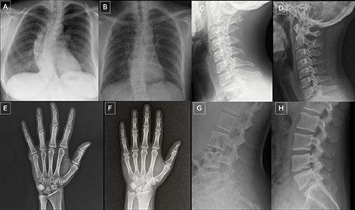
Anatomy-matched real and GPT-4o-generated radiographs: (A) real and (B) GPT-4o-generated posteroanterior chest radiographs, (C) real and (D) GPT-4ogenerated lateral cervical spine radiographs, (E) real and (F) GPT-4o-generated posteroanterior hand radiographs, and (G) real and (H) GPT-4o-generated lateral lumbar spine radiographs. The pairs demonstrate that GPT-4o can produce radiographically plausible images across different anatomic regions.